ACUTE OTITIS MEDIA
I Made Nudi Arthana
Acute otitis media , a inflammation ear middle relate with effusion ear middle , which is accumulation fluid in the ear middle . Otorrhea is an ear discharge that can originate from tympanic membrane . Otitis media is classified based on symptom clinical , otoscopy , duration of illness and complications . Otitis media occurs Because aeration ear disturbed middle , usually due to Because impaired eustachian tube function . Correct diagnosis and management very much important , because otitis media is common diseases found and can cause complications distribution infection until to intracranial. 3
LITERATURE REVIEW
Anatomy Ear
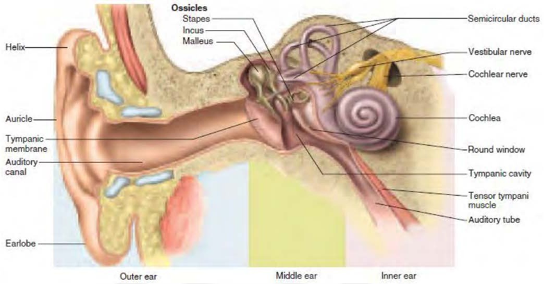
Picture 1. Anatomy ear.
Outer Ear
Ear outside consists of from leaf ear (auricle), hole ear (meat) external acusticus) to the lateral tympanic membrane. The auricle is formed by cartilage and muscle and covered by skin. Towards the ear canal lining funnel-shaped cartilage covers almost one-third of the lateral, two-thirds other hole ear formed by bone Which covered skin Which attached close And relate with membrane timpani. Form leaf ear with various bulge And basin as well as form hole ear Which straight with long around 2.5 cm, will cause a sound resonance of 3500 Hz. One third The outer part consists of cartilage which contains many cerumen glands and hair, while the inner two-thirds consist of bone with a little earwax. 5
Middle Ear
The middle ear is cube-shaped and consists of the tympanic membrane, the tympanic cavity, and the tympanic membrane, eustachian tube, and auditory ossicles. The upper part of the tympanic membrane called the pars flaccida (Shrapnell's membrane) which consists of two layers, namely layer outside is advanced epithelium skin hole ear And layer in coated by cell cube ciliated. Part lower membrane timpani called pars tension (membrane propria) Which have one layer in middle, namely layer Which consists of from fiber collagen And A little fiber elastin. Bone hearing consists of on malleus, the incus, and stapes are arranged from the outside to the inside like a connected chain from membrane timpani going to cavity ear in. Process long malleus attached to the tympanic membrane, the malleus is attached to the incus, and the incus is attached to the on the stapes. The eustachian tube connects the nasopharynx to the middle ear. middle. Process mastoid is part bone temporalis Which located in behind the ear. The air space above it is called the antrum. mastoid Which relate with cavity ear middle. Infection can spreads from the middle ear cavity to the mastoid antrum which can cause mastoiditis. 4
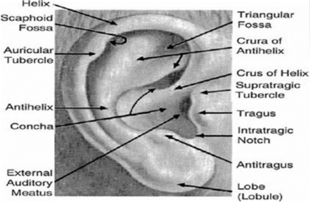
Picture 2. Auricle. 6
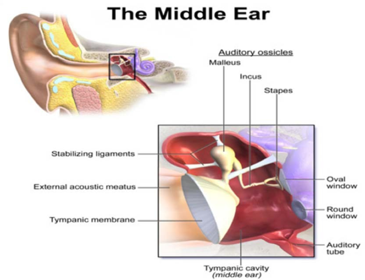
Picture 3. Ear middle. 6
Inner Ear
Ear in consists of from two part, that is labyrinth bone And labyrinth membranous. Labyrinth bone consists of from cochlea, vestibule, And canal semi- circular, while the membranous labyrinth consists of the utricle, saccule, ductus cochlear, and semicircular ducts. The cavity of the bony labyrinth is lined by a layer thin internal periosteum or endosteum, and is mostly filled by trabeculae (the arrangement resembles a sponge). The cochlea (cochlea) is two and a half in shape circle. End or peak cochlea called helicotrema, connect perilymph scale vestibule (adjacent on) And scale timpani (adjacent lower). Between vestibular scale and tympani scale contain the media scale (cochlear duct). vestibule And scale timpani containing perilymph with 139 mEq/l, whereas scale
media containing endolymph with 144 mEq/l mEq/l. Matter This important For hearing. Base scale vestibule called membrane vestibular ( Reissner's Membrane ) whereas base scale media is membrane basilar. On This membrane contains the organ of Corti which contains important organelles for mechanism nerve peripheral hearing. Organ Corti consists of from One line cell hair inner row containing 3,000 cells and three rows of outer hair cells containing 12,000 cells. The afferent and efferent nerve endings are attached to the lower end of the hair cells. surface cells hair there is stereocilia Which attached on a sheath in above it Which tend flat, known as membrane tectoria. Membrane tectoria secreted And supported by a stage Which located in medial called as limbus. 5
The auditory nerve or hearing nerve consists of two parts, namely: vestibular nerve (balance) and cochlear nerve (hearing). The fibers fiber nerve vestibular move going to nucleus vestibular Which is at on point meeting between punch And medulla oblong, Then going to cerebellum. Whereas, fiber nerve nerve cochlear at first emitted to A nucleus special Which is at appropriate in behind thalamus, Then emitted Again towards the final receiving center in the cortex of the brain located at the bottom temporal lobe. The vascularization of the inner ear originates from the A. Labyrinthine branch of A. Cerebellaris anteroinferior or branch from A. Basilaris or A. Vertebral. Artery This enter to meatus internal acoustics And separated become A. Vestibular front And A. Cochlear communism Which branching also become A. Cochlear And
A. Vestibulocochlear. A. Vestibular anterior supplies N. Vestibularis, urticulus and part of the semicircular ducts. A. Vestibulocochlearis reaches the middle area round basal cochlea separated become branch terminal vestibular and the cochlear branch. The vestibular branch supplies the saccule, most of which canal semicircular And end basal cochlea. Branch kohlear to bleed spiral ganglion, spiral lamina ossea, limbus and spiral ligament. A. Cochlear runs around the N. Acousticus in the internal acoustic canal and in the cochlea around the modiolus. The veins drain into the V. Labyrinthine which continues into the sinus inferior petrosal or sigmoid sinus. Small veins pass through the aqueduct vestibular And cochlear to sine petrosus superior And inferior. Nerves ear through the N. Vestibulocochlearis (N. acousticus) which is formed by part of cochlear and vestibular, in the internal acoustic meatus unite on the lateral side the root of the facial nerve and enters the brain stem between the pons and medulla. Sensory cells The vestibular nerve is innervated by the N. Cochlearis with the vestibular ganglion (Scarpa) located at the bottom from meatus acoustician internal. Cells sensory hearing innervated N. Cochlear with ganglion spiral corti located in modiolus. 5
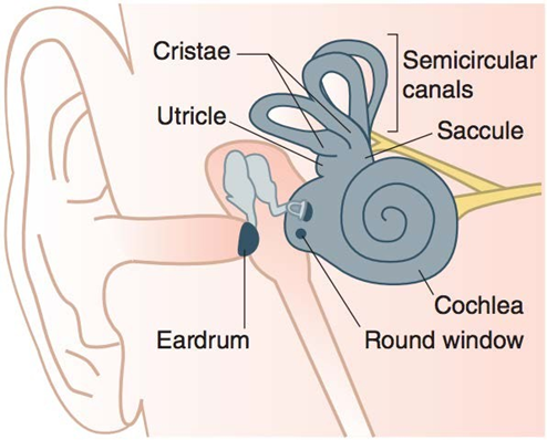
Picture 4. Inner ear . 7
Ear Physiology
The hearing process begins with the sound energy being captured by the leaves. ear in form wave Which streamed through air or bone to cochlea. The vibrations vibrate the tympanic membrane and are transmitted to the ear. middle through a series of auditory ossicles which will imply vibration through Power leverage bone hearing And multiplication comparison wide membrane timpani And window oval. Energy vibration Which has amplified This will passed to the stapes which moves the oval window so that the perilymph is in scale movable vestibule.
Vibration forwarded through membrane Reissner Which pushing the endolymph, so that it will cause relative movement between the basilar membrane and the membrane tectoria. This process is a mechanical stimulus that causes the occurrence of deflection stereocilia cells hair, so that channel ion open And happen release ion loaded electricity from body cell. Condition This cause process depolarization of hair cells, thereby releasing neurotransmitters into the synapse which will cause an action potential in the auditory nerve, then continue to nucleus auditory until to cortex hearing (area 39-40) in lobe temporalis. 8
Factors Risk
Otitis media is disease multifactorial , several the most frequent factor influence the occurrence of acute otitis media (AOM) is infection , allergies , and environment . 1
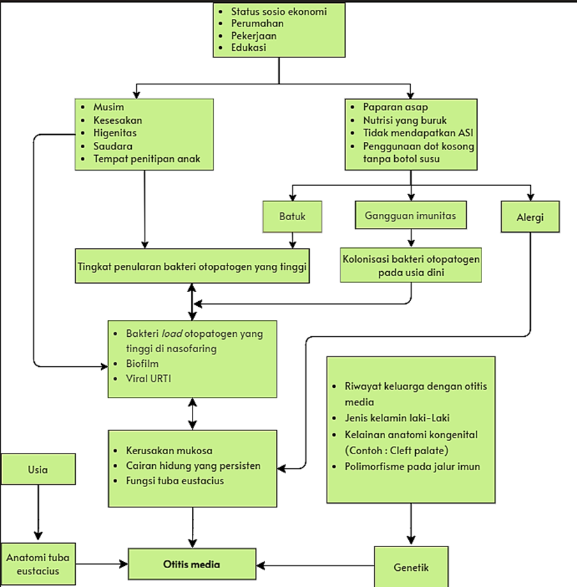
Figure 5. Causal and Factor Flow OMA risk . 11
Condition infection channel breathing on is factor the most common risks the occurrence of AOM in children can caused by viruses or bacteria . Bacteria pathogens , such as Streptococcus pneumoniae , Haemophilus influenza , and Moraxella ( Branhamella ) catarrhalis responsible answer on more of 95% of cases . While Viral pathogens such as respiratory syncytial virus , influenza virus, parainfluenza virus, rhinovirus, and adenovirus are also commonly developing OMA. 3 Otitis media begins as an inflammatory process after respiratory tract viral infection breathing above which involves mucosa nose , nasopharynx , mucosa ear middle , and Eustachian tube . Because the space anatomy ear narrowed center , edema caused by the inflammatory process obstruct part narrowest from the Eustachian tube which then cause decline ventilation . This is cause a series incident that resulted in improvement pressure negative in the ear middle , increase exudate from inflamed mucosa , and accumulation of secretion mucosa , which allows colonization organism bacteria and viruses in the ear middle . Growth microbes this is in the ear middle Then produce pus in the space ear middle 15 . This is shown in a way clinically by a protruding tympanic membrane or erythematous and fluid ear purulent center . This must differentiated from suppurative otitis media chronic (CSOM), which appears with fluid thick colored yellow in the room ear middle and retraction tympanic membrane (TM) on examination otoscopy . Both will produce decline MT mobility in tympanometry or pneumatic otoscopy . 11
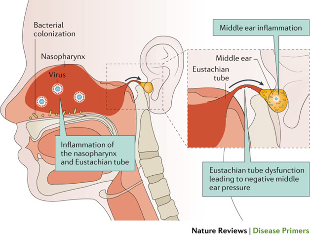
Figure 6. Pathogenesis of Virus- Induced AOM . 11
Temporary factor risk other like type sex male , adenoid hypertrophy ( obstruction ), allergies , vitamin A deficiency , abnormalities anatomy palatum and tensor veli palatini, dysfunction cilia , as well as implant the cochlea also affects occurrence of OMA. Visit to place custody child , social status economy low , exposure to passive smoke in the environment , lack breastfeeding , pacifier use , deficiency immune ( human immunodeficiency virus /HIV, diabetes, and iron deficiency) immune others ), reflux gastroesophageal , history family with recurrent AOM in the elderly or you womb , and predisposition genetics other is factor other risks of OMA. Mucins are included abnormality expression of this gene , especially MUC5B upregulation is also known related with OMA. 1
Management
Journey acute otitis media (AOM) disease can healed with Good in a way natural in healthy children with symptom settle down in a number of day and rarely happen complications , although without therapy antibiotics . Besides That use routine antibiotics in condition general like OMA also increases risk resistance antimicrobial , both at the level of community and also individual , so that risks and benefits This must under consideration with Good customized with patient clinical . 6 For child Healthy aged ≥6 months who have disease light with the correct diagnostic criteria for AOM or child who is not fully fulfil criteria diagnostics , observation for 48 hours with giving analgesic recommended , and recommended for . 6 :
Evaluate repeat child in 24 hours to 48 hours for documenting journey clinical ; or
Request caregiver or parents patient return If child No get better or getting worse anytime in 48 hours; or
Give recipe antimicrobial For consumed If child No get better .
Giving antibiotics said to be most effective in children age not enough from 2 years with bilateral AOM and in all age experienced Secret ear I due to OMA. So , the child with condition This recommended For giving antibiotics quick based on latest guidelines . 6 Treatment antibiotics quick recommended for OMA in patients age not enough than 6 months , immunocompromised , or own malformation craniofacial , as well as they with disease heavy due to OMA. In children with OMA no complex and not weight that is not at risk tall experience complications , can done observation and prescription antibiotics can postponed , unless persistent OMA symptoms for 48-72 hours. 8
Therapy Pharmacological
Once the diagnosis of acute otitis media (AOM) is established , the goal the therapy
is For therapy symptomatic including control pain and symptoms systemic in the form of fever , and treat the infection process with antibiotics . Therapy pharmacological in the form of drug nonsteroidal anti-inflammatory drugs (NSAIDs, such as ibuprofen) or acetaminophen ( paracetamol ) can used For control pain . Paracetamol can given 10 to 15 mg/ kgBW every 4 to 6 hours in children , and 500 mg every 4 to 6 hours in adults. (Uum et al., 2019) . There is controversy about prescription antibiotics in early otitis media as explained previously . Giving antibiotics indicated in the condition certain , such as children with MT protrusion accompanied by fever (≥39°C) and complaints systemic currently until severe , or who have experienced severe otalgia , or who have Sick heavy for 48 hours. In addition that , when suspected existence etiology bacteria as in suppurative stage OMA , administration of oral antibiotics can recommended . Therapy line first in patients who do not allergy to penicillin is amoxicillin dose high in patients children and adults 2 :
Amoxicillin 75 mg/ kgBW / day up to 90 mg/ kgBW / day shared twice a day in stock capsule or suspension ; or
Amoxicillin 45 mg/ kgBW / day up to 60 mg/ kgBW / day shared three times a day in stock capsule or suspension .
Giving antibiotics dose appropriate for five days recommended For
most child aged ≥2 years with OMA without complications , whereas giving antibiotics during ten day recommended For more children young ( six) up to 23 months ) and cases with MT perforation or recurrent OMA 2 . In case allergy penicillin , the American Academy of Pediatrics (AAP) recommends azithromycin dose single 10 mg/ kgBW or clarithromycin (15 mg/ kgBW per day ) in 2 doses divided ). Another option for patient allergy penicillin is cefdinir 14 mg/ kgBW per day in 1 or 2 doses , cefpodoxime 10 mg/ kgBW per day , once a day a day , or cefuroxime 30 mg/ kgBW per day in 2 doses divided 11. Oral antibiotics reduce duration symptoms of AOM and effusion ear middle in succession , but need be noticed effect possible side arise , such as gastrointestinal symptoms and rash skin . If there is any MT perforation , therapy must to be continued with antibiotics safe ototopical For use in the ear middle , such as ofloxacin, than antibiotics systemic , because This give concentration far away antibiotics more tall without effect systemic side . 2
If recurrent AOM occurs , management will focuses on preventing further AOM episodes continue . Immunization with PCV in infancy beginning has proven effective in reduce risk child develop recurrent OMA , however No effective For more children big . Prophylaxis antibiotics in children with recurrent OMA reduce amount OMA recurrence from 3 recurrences to 1.5 per year . However , its use prolonged No recommended remember effect side effects and antibiotic resistance 18 . For patients whose symptoms No get better after treatment with amoxicillin dose high , amoxicillin-clavulanate dose tall with 90 mg/ kgBW per day component amoxicillin , and 6.4 mg/ kgBW per day clavulanate in 2 doses divided must given . In children who vomit or If There is situations where oral antibiotics are not can given , ceftriaxone 50 mg/ kgBW per day during three day in a row , good in a way intravenous or intramuscular is choice 16
Decongestant topical and oral, antihistamines and corticosteroids Not yet proven effective or has show conflicting results in overcome OMA symptoms and therefore That No recommended . Tympanocentesis or myringotomy , incision small from tympanic membrane which allows fluid flow from ear middle , maybe own role in determine pathogen causes of OMA, but No effective as modality treatment for OMA 8 .
Non -Pharmacological Therapy ( Surgery )
Procedure tympanocentesis is one of action For reduce pressure cavity ear in the middle of the OMA case . Actions taken with enter needle small to in tympanic membrane , expanded to cavity ear middle , and take accumulated fluid . Procedure This effective for diagnostics and to provide benefit therapeutic in reduce pain and pressure ear middle Because drainage fluids , as well as can increase delivery agent antibiotics topical to cavity ear middle infected 6,7,10 .
Patients who have experience four or more OMA episodes in two twelve month final must under consideration For myringotomy with placement tube ventilation ( grommets ), according to guidelines American Academy of Pediatrics 11 . Recommendations practice clinical from American Academy of Otolaryngology Head and Neck Surgery moment This also recommends installation tube tympanostomy in children with recurrent AOM and effusion at the time of evaluation 8 . Tube ventilation ( tympanostomy or myringotomy ) is tube plastic small inserted to in tympanic membrane (TM) through operation short with anesthesia general and also local . Tube will placed for 6 to 12 months and can off alone . Indications main action surgery This is recovery hearing in children with chronic otitis media with effusion ( glue ear ) and prevention recurrence in children with recurrent AOM with emit fluid from ears and improve ventilation . Besides that , with give access to ear middle , tube ventilation allow For treatment antibiotics topical for OMA. Benefits tube ventilation especially for 6 months First after insertion has documented , with around one episode of OMA can prevented . Although No definitive , proof moment This about journey natural and benefits treatment show that tube ventilation No effective for recurrent OMA without effusion ear persistent middle , but is the right choice For managing recurring OMA with effusion ear middle persistent in one or both ears 11.15 . Tube tympanostomy increase quality life specific disease in patients , although Not yet proven significant in lower number recurrence of AOM. Increased big in field psychosocial suffering physical , disorders hearing , disorders talk , pressure emotional , and limitations activity documented after action tympanostomy . Installation tube tympanostomy No related with complications main like disturbance sensorineural hearing loss , bleeding consequence injury vascular , and termination chain bone hearing . However can happen a number of complications general including drainage ears , good ejection or preservation inserted tube , formation inflammation benign , and residual perforation 8 .
Myringoplasty required If perforation happen together with infection secondary and interference hearing . Tube tympanostomy can migrate from the location in the ear canal and moves to behind intact tympanic membrane in complications asymptomatic which is rare 11 . Other structures such as adenoids that function as a nasopharyngeal reservoir pathogen breathing , when grow bigger can cause obstruction nasal airway and interferes with Eustachian tube function . Adenoidectomy is operation Adenoid removal performed on children with recurrent OMA For increase function ear middle and with thus prevent further episodes of AOM 11 . However do adenoidectomy just No recommended For prevention of OMA, must still under consideration installation tube tympanostomy Because more beneficial . Chemoprophylaxis with amoxicillin dose low show decline significant frequency of OMA compared to with placebo in two study with a period of action carry on One year . Intervention surgery must under consideration in recurrent OMA cases , both Because Eustachian tube dysfunction or resistance bacteria , Down syndrome , and abnormalities craniofacial like cleft palate 8.9
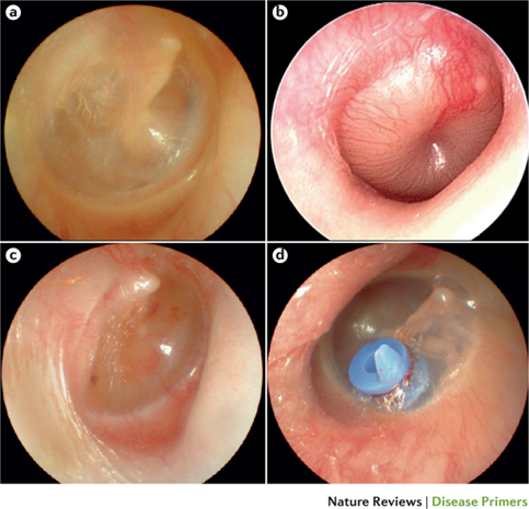
Figure 7. Condition Tympanic Membrane . (a) normal MT; (b) hyperemic and prominent MT. indicates OMA; (c) otitis media with effusion ; (d) MT installed tube tympanostomy 11 .
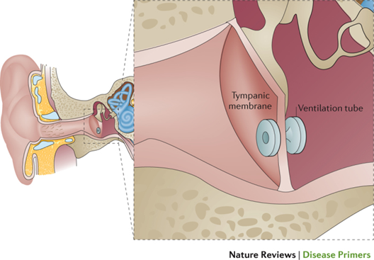
Figure 8. Installation Tube Tympanostomy at MT 10 .
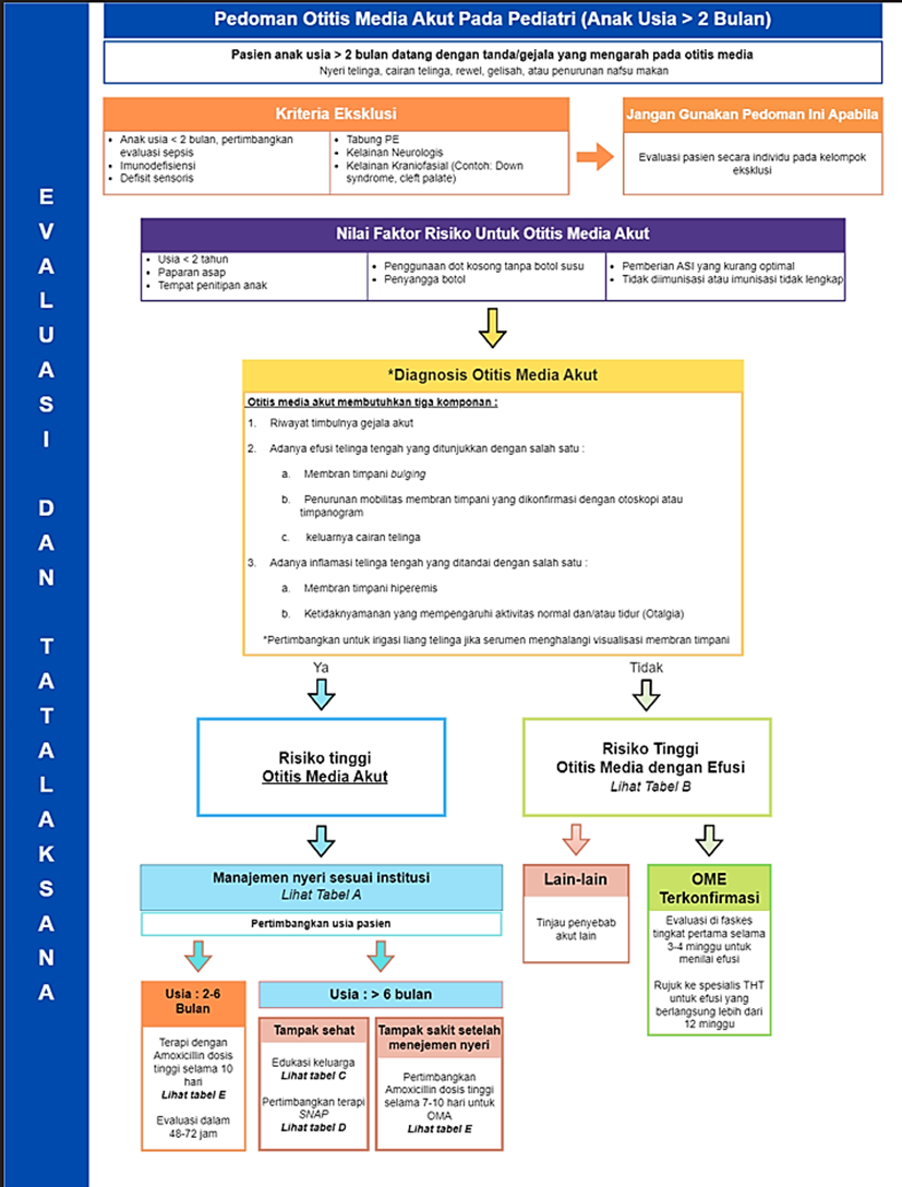
Figure 9. OMA Management Chart 7
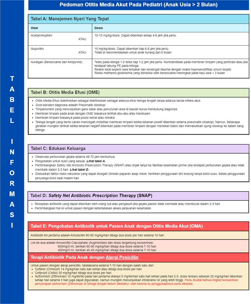
Figure 10. OMA Management Chart ( continued ) 7 .
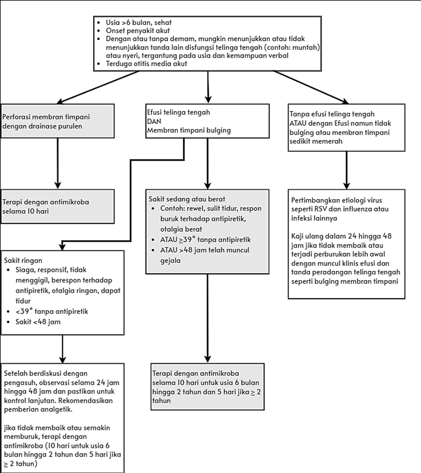
Figure 11. SummaryManagement of OMA 10 .
Conclusion
otitis media is inflammation ear middle caused by infection channel breathing acute . The virus will enter , causing inflammation and damage protector experience from ear allow the occurrence infection bacteria.pain Alone caused by inflammatory processes caused by viruses and bacteria , which cause gathering fluid in the ear part middle so that tympanic membrane will looks stands out , and when happen perforation will cause the exit fluid from ear . pain in this OMA related with inflammatory process in the ear
Where some large OMA in children started with existence infection in the tract breath part above . the virus causes inflammation of the nasopharynx and disorders function from the Eustachian tube . Eustachian tube Alone is protector natural that prevents colonization from nasopharynx to ear middle . Children usually prone to against acute otitis media Because immunity systemic that is not mature and more horizontal tube anatomy . Viruses/ bacteria bother cleaning mucociliary and causes blockage of the Eustachian tube , so that pressure negative occurs in the ear middle . Pressure negative This facilitate entry pathogenic bacteria and viruses to in cavity ear middle cause inflammation ear middle , accumulation fluid ear middle ear , and symptoms of acute otitis media . The most common symptoms of AOM in adults complained about is painful sudden ear , but in children who have not Can talk usually marked with hold ear , crying excess , fever , disturbance sleep . Examination otoscope in acute otitis media can be found changes in the tympanic membrane such as hyperemia of the tympanic membrane or bulging was found on the tympanic membrane . In children , acute otitis media can diagnosed If found fluid in the ear middle and accompanied complaint fever , sick ears , irritability , concomitant with symptom disturbance system breathing Acute otitis media (AOM) is greatly influenced by environmental factors . risk in the form of factor often experience ISPA disease and the therapy that must be given right so as not to occurrence of suppurative otitis media chronic (CSOM) type dangerous can result in fatal complications . It has been reported by WHO that disease channel breath on is reason most frequent the occurrence of acute otitis media (AOM).
REFERENCE
Ardıç, C., & Yavuz, E. (2018). Effect of breastfeeding on common pediatrics infections: A 5-year prospective cohort study. Archivos Argentinos de Pediatrics , 116 (2), 126–132. https://doi.org/10.5546/aap.2018.eng.126
2. Chiappini, E., Ciarcià, M., Bortone, B., Doria, M., Becherucci, P., Marseglia, G.L., Motisi, M.A., de Martino, M., Galli, L., Licari, A. , De Masi, S., Lubrano, R., Bettinelli, M., Vicini, C., Felisati, G., Villani, A., & Marchisio, P. (2019). Updated Guidelines for the Management of Acute Otitis Media in Children by the Italian Society of Pediatrics: Diagnosis. The Pediatric Infectious Disease Journal , 38 (12S), 3–9. https://doi.org/10.1097/INF.0000000000002429
3. Chonmaitree, T., Jennings, K., Golovko, G., Khanipov, K., Pimenova, M., Patel, J. A., McCormick, D. P., Loeffelholz, M. J., & Fofanov, Y. (2017). Nasopharyngeal microbiota in infants and changes during viral upper respiratory tract infection and acute otitis media. PLOS ONE , 12 (7), 1–15. https://doi.org/10.1371/journal.pone.0180630
4. Folino, F., Bosi, P., Torretta, S., Gaffuri, M., & Marchisio, P. (2022). Recurrent Acute Otitis Media Environmental Risk Factors : A Literature Review from the Microbiota Point of View. Appl. Microbiol. , 2 , 594–613.
5. Hassooni, HR, Fadhil, SF, Hameed, RM, Alhusseiny, AH, & Ali Jadoo, SA (2018). Upper respiratory tract infection and otitis media are clinically and microbiologically associated. Journal of Ideas in Health , 1 (1), 29–33. https://doi.org/10.47108/jidhealth.vol1.iss1.7
6. Hayashi, T., Kitamura, K., Hashimoto, S., Hotomi, M., Kojima, H., Kudo, F., Maruyama, Y., Sawada, S., Taiji, H., Takahashi, G ., Takahashi, H., Uno, Y., & Yano, H. (2020). Clinical practice guidelines for the diagnosis and management of acute otitis media in children—2018 update. Auris Nasus Larynx , 47 (4), 493–526. https://doi.org/10.1016/j.anl.2020.05.019
7. Huntington, M. K., VanKeulen, S., & Hoffman, W. W. (2013). What Ever Happened to the Common Cold? Improving Antibiotic Utilization. South Dakota Medicine , 136–146.
8. Jamal, A., Alsabea, A., Tarakmeh, M., & Safar, A. (2022). Etiology, Diagnosis, Complications, and Management of Acute Otitis Media in Children. Cureus , 14 (8), 1–7. https://doi.org/10.7759/cureus.28019
9. Lahiry, S., Dalal, K., Pawar, A., & Kotak, B. (2021). High-Dose amoxicillin supported with clavulanic acid as empirical therapy in acute otitis media. Asian Journal of Medical Sciences , 12 (4), 134–144. https://doi.org/10.3126/ajms.v12i4.33541
10. Lieberthal, AS, Carroll, AE, Chonmaitree, T., Ganiats, TG, Hoberman, A., Jackson, MA, Joffe, MD, Miller, DT, Rosenfeld, RM, Sevilla, XD, Schwartz, RH, Thomas, PA, & Tunkel, DE (2013). The Diagnosis and Management of Acute Otitis Media. American Academy of Pediatrics , 131 (3), 964–999.
11. Marchisio, P., Bortone, B., Ciarcià, M., Motisi, M.A., Torretta, S., Gattinara, G.C., Picca, M., Mauro, G. Di, Bonino, M., Mansi, N. , Varricchio, A., Marseglia, G.L., Cardinale, F., Villani, A., & Chiappini, E. (2019). Updated Guidelines for the Management of Acute Otitis Media in Children by the Italian Society of Pediatrics. The Pediatric Infectious Disease Journal , 38 (12S), 22–36. https://doi.org/10.1097/INF.0000000000002430
12. Martines, F., Salvago, P., Ferrara, S., Messina, G., Mucia, M., Plescia, F., & Sireci, F. (2016). Factors influencing the development of otitis media among Sicilian children affected by upper respiratory tract infections. Brazilian Journal of Otorhinolaryngology , 82 (2), 215–222. https://doi.org/10.1016/j.bjorl.2015.04.002
13. Mittal, R., Robalino, G., Gerring, R., Chan, B., Yan, D., Grati, M., & Liu, X. Z. (2014). Immunity genes and susceptibility to otitis media: A comprehensive review. Journal of Genetics and Genomics , 41 (11), 567–581. https://doi.org/10.1016/j.jgg.2014.10.003
14. Ngo, CC, Massa, HM, McMonagle, BA, Perry, CF, Nissen, MD, Sloots, TP, Thornton, RB, & Cripps, AW (2022). Predominant Bacterial and Viral Otopathogens Identified Within the Respiratory Tract and Middle Ear of Urban Australian Children Experiencing Otitis Media Are Diversely Distributed. Frontiers in Cellular and Infection Microbiology , 12 (775535), 1–11. https://doi.org/10.3389/fcimb.2022.775535
15. Rosenfeld, RM, Tunkel, DE, Schwartz, SR, Anne, S., Bishop, CE, Chelius, DC, Hackell, J., Hunter, LL, Keppel, KL, Kim, AH, Kim, TW, Levine, JM, Maksimoski, MT, Moore, DJ, Preciado, DA, Raol, NP, Vaughan, WK, Walker, EA, & Slp, C. (2022). Clinical Practice Guideline: Tympanostomy Tubes in Children (Update). Otolaryngology–Head and Neck Surgery , 166 (1S), 1–45. https://doi.org/10.1177/01945998211065662
16. Saux, N. Le, Robinson, J. L., & Society, C. P. (2016). Canadian Pediatric Society Position Statement: Management of acute otitis media in children six months of age and older. Pediatric Child Health , 21 (1), 39–44.
17. Schilder, A.G.M., Chonmaitree, T., Cripps, A.W., Rosenfeld, R.M., Casselbrant, M.L., Haggard, M.P., & Venekamp, R.P. (2016). Otitis media. Nature Reviews Disease Primers , 2 , 1–19. https://doi.org/10.1038/nrdp.2016.63
18. Seppälä, E., Sillanpää, S., Nurminen, N., Huhtala, H., Toppari, J., Ilonen, J., Veijola, R., Knip, M., Silifä, M., Laranne, J ., Oikarinen, S., & Hyöty, H. (2016). Human enterovirus and rhinovirus infections were associated with otitis media in a prospective birth cohort study. Journal of Clinical Virology , 85 , 1–6. https://doi.org/10.1016/j.jcv.2016.10.010
19. Uum, RT Van, Venekamp, R.P., Schilder, AGM, Damoiseaux, RAMJ, & Anthierens, S. (2019). Pain management in acute otitis media: a qualitative study of parents' views and expectations. BMC Family Practice , 20 (18), 1–7.
20. Vila, P. M., Ghogomu, N. T., Odom-John, A. R., Hullar, T. E., & Hirose, K. (2018). Infectious complications of pediatric cochlear implants are highly influenced by otitis media. Physiology & Behavior , 97 , 76–82. https://doi.org/10.1016/j.ijporl.2017.02.026
21. Zernotti, M.E., Pawankar, R., Ansotegui, I., Badellino, H., Croce, J.S., Hossny, E., Ebisawa, M., Rosario, N., Sanchez Borges, M., Zhang, Y. , & Zhang, L. (2017). Otitis media with effusion and atopy: Is there a causal relationship? World Allergy Organization Journal , 10 (37), 1–9. https://doi.org/10.1186/s40413-017-0168-x
Comments