ANTROCHOCANAL POLYP AND INFERIOR CONCHA HYPERTROPHY
I Made Nudi Arthana
Antrochoanal polyp is a benign polypoid lesion, originating from the inflamed and edematous mucosa of the maxillary antrum, consisting of a cystic part in the intra-antrum and a solid polypoid part in the intra-nasal region characterized by swelling of the mucosal mass. Which inflamed with stalk, Which Then go out from ostium sine and expand until to behind in area koana posterior. Most polyp originate from the osteomeatal cleft causing nasal obstruction. Polyps often grow in the ethmoidal and maxillary sinuses. These polyps are also known as Killian's polyps because they were first discovered by Killian in 1906. 1
LITERATURE REVIEW
Anatomy of the nose and paranasal sinuses
Nose consists of on nose outside And nose in. Nose outside formed by bone hard And bone vulnerable, network tie as well as muscles small Which functioning to widen hole nose. Nose in consists of on structure Which stretch from the os internum anteriorly to the choana posteriorly, which separates the nasal cavity from the nasopharynx. The nasal cavity is tunnel-shaped from front to back, separated by septum rice in the the middle become cavity rice right And left. The vestibule is a part of the nasal cavity that is located in the ala nasi, just behind the anterior nares. The vestibule is covered by skin that has many sebaceous glands and long hairs called vibrissae. 7-8
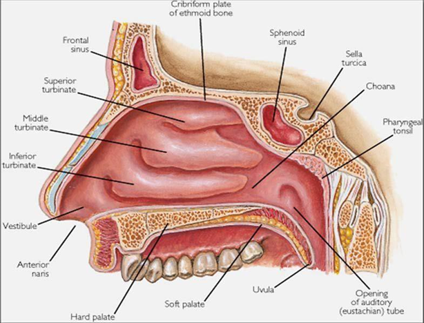
Picture 1. Anatomy nose 9
Each nasal cavity is limited by 4 walls, namely the medial, lateral, inferior and superior walls. The medial wall of the nose is the nasal septum. This nasal septum is formed by bone and cartilage, the lateral wall contains the superior concha, middle concha and inferior concha and the supraconcha is usually rudimentary. The inferior concha is a separate bone that is attached to the maxillary bone and the ethmoid labyrinth, while the medial, superior and supraconcha are part of the labyrinth ethmoid. Gap between the inferior concha and the base of the nose is called the inferior meatus, next gap between concha media And inferior called meatus media and above the middle concha is called the superior meatus. 7,9
The upper part of the nasal cavity receives blood from the anterior ethmoid artery. And posterior Which is branch from a. ophthalmic from a.carotid internal. Section lower cavity nose get bleeding from branch a. maxillary internal, including the tips of the greater palatine artery and the sphenopalatine artery which exit the sphenopalatine foramen with the sphenopalatine nerve and enter the nasal cavity behind the posterior end of the middle concha. The front of the nose receives blood from branch – branch a.facialist. On part front septum there is anastomosis from branches of the sphenopalatine artery, anterior ethmoid artery, superior labial artery, and greater palatine artery called the Kiesselbach plexus (Little's area). The veins of the nose have the same name and run side by side with the arteries. The veins in the vestibule and the outer structures of the nose drain into the ophthalmic vein which is connected to the cavernous sinus. The veins in the nose do not have valves, thus predisposing to easy spread of infection to the intracranial. 7.10
The nose is innervated by sensory nerves derived primarily from the ophthalmic and maxillary branches of the trigeminal nerve. The rest of the nasal cavity receives sensory innervation mostly from the maxillary nerve via the sphenopalatine ganglion. The olfactory nerve descends through the lamina cribrosa from the undersurface of the bulb. olfactory And then spread across mucosa lining the upper part of the superior concha and the superior part of the septum. 8
Sine paranasalis is cavities Which there is in bone maxillary, frontal, sphenoid And ethmoidalis. There is four install sine paranasal, four on each side of the nose; right and left frontal sinuses, ethmoidal sinuses right And left (anterior And posterior), sine maxillary right And left and the right and left sphenoid sinuses. Clinically the paranasal sinuses are divided into two groups that is part front And posterior. Group front culminate in below the middle concha, at or near the infundibulum, consists of the frontal sinus, maxillary sinus, And cells front sine ethmoid. Group posterior culminate in various places above the middle concha consist of posterior cells of the ethmoid sinus and sphenoid sinus. 10
The maxillary sinus is the largest paranasal sinus. It is estimated that the formation of the sinus occurs on the 70th day of pregnancy. At birth the volume of the maxillary sinus is around 6-8 ml, then it grows rapidly and finally reaches size maximum that is 15 ml on moment mature. Sine maxillary empties into in the middle meatus through the hiatus semilunaris. Bleeding obtained from the branch artery maxillary internal including infraorbital, branch lateral sphenopalatine, anterior superior and posterior alveolar arteries. Most of the maxillary sinus wall is drained by the maxillary vein which is connected to the pterygoid plexus. Mucosa sine maxillary served nerve alveolar superior And infraorbital nerve. The accessory bone of the maxillary sinus is located anterior to the uncinate process. 8,11
The sphenoid sinuses are formed in the 3-month-old fetus as paired mucosal evaginations in the posterior superior cavity rice. Before the child is 3 years old, the sphenoid sinus is still small, but has fully developed at the age of 12 to 15 years. It is located in the corpus of the ethmoid bone and its size and shape vary. This pair of sinuses is separated from each other by a thin bony septum, which is rarely located right on middle, so that Wrong One sine will bigger than the other side. Location OS sphenoid is in in sphenoid bone in behind the posterior ethmoid sinus. The sphenoid sinus is divided into two by a partition called the intersphenoid septum. The size is tall 2 cm, inside 2.3 cm, And width 1.7 cm. The volume ranges from 5 to 7.5 ml. 8-9

Picture 2. Anatomy sine paranasalis 9
Sine ethmoid is sinus that can be a focus of infection for other sinuses. The ethmoid cells, first formed in the 4-month-old fetus, originate from meatus superior And supremacy Which to form group cells Anterior and posterior ethmoid. The ethmoid sinuses are already present at birth and then develop according to age until they reach puberty. In adults, the shape of the ethmoid sinuses is like a pyramid with its base at posterior part. Its size is from anterior to posterior 4-5 cm, height 2.4 cm, and 0.5 cm wide anteriorly and 1.5 cm posteriorly, the volume of the sinus is approximately 14 ml. The ethmoid sinus is hollow – the cavity consists of cells that resemble a wasp nest, Which there is in in mass part lateral OS ethmoid, Which located in between media konka and the medial wall of the orbit. Based on its location, the ethmoid sinus is divided into the anterior ethmoid sinus which empties into the middle meatus, and the posterior ethmoid sinus Which culminate in meatus superior. In part at the forefront sine ethmoid Anteriorly there is a narrow section, called the frontal recess, which is connected to the frontal sinus. The largest ethmoid cells are called ethmoid bullae. In the anterior ethmoid region there is a narrowing of the infundibulum, where the maxillary sinus ostium opens. Swelling or inflammation in the frontal recess can cause frontal sinusitis and swelling in the infundibulum can cause maxillary sinusitis. Roof sine ethmoid Which called fovea ethmoidalis bordering with lamina cribrosa. The lateral wall of the sinus is a very thin lamina papyracea that lines the sine ethmoidalis from the cavity orbit. In part behind sine The posterior ethmoidal bone borders the sphenoid sinus. 7-10
The shape and size of the frontal sinuses vary greatly and sometimes there are rudimentary sinuses. The shape of the right and left frontal sinuses is usually not symmetrical, One more big from on other And separated by partition Which located in the midline. Approximately 15% of adults have only one frontal sinus and approximately 5% of their frontal sinuses are underdeveloped. The average size of the frontal sinus: 3 cm high, width 2-2.5 cm, in 1.5-2 cm, and the average content is 6-7 ml. The absence of septum or sinus wall concavities on X-rays indicates existence infection sine. Sine frontal separated by bone Which relatively thin from the orbit and anterior cerebral fossa, so that infection from the frontal sinus can easily spread to area This. Sine frontal drained through the ostium Which located in the frontal recess which is related to the ethmoid infundibulum. 8,10
Function Nose and Paranasal Sinuses
Based on structural theory, revolutionary theory and functional theory, the physiological functions of the nose are:
Function respiration For arrange condition air ( water conditioning ), filter air, humidification, balancing in pressure exchange and local immunological mechanisms
Function smell Because there is mucosa olfactory (smell) And air reservoir to accommodate olfactory stimuli
Function phonetics Which useful For resonance voice, help process speak and prevent the conduction of sound itself through bone conduction
Statistical and mechanical functions to relieve head load, protection against trauma and heat protection
reflex . 25
The three main functions of the nose as olfactory, respiratory and protective functions are supported by the anatomy of the nasal cavity, which requires a large surface area. Which wide. Condition from mucosa nose, humidity as well as surface from the ciliated nasal cavity increases contact with inspired air, which can maximize function smell And warm, moisturize And filtering incoming air before it reaches the lower respiratory tract. The presence of good vascularization and the secretory system of the nasal cavity and paranasal sinuses causes air inspiration become warm And moist, before reach channel breath part lower. The existence of flow turbulence air on nose is The main physiology of the nose. Turbulent flow of air in the nasal cavity increases contact between inspired air and the nasal mucosa, which plays a role not only in respiratory function but also in olfactory function and body defense. 24
Factors that play a role in maintaining the function of the paranasal sinuses are patency. COM, function transportation mucociliary And production mucus Which normal. KOM Patency own role Which important as place drainage mucus And debris and maintain oxygen pressure in condition normal thus preventing the growth of bacteria. Mucociliary transport factors are highly dependent on the characteristics of the cilia, namely the structure, number and coordination of cilia movement. Mucus production also depends on the volume and viscoelasticity of mucus which can affect mucociliary transport. 25
A number of theory Which put forward as function sine paranasal between others are:
As regulator air condition ( air conditioning )
As retainer temperature ( thermal insulators )
Help balance head
Help sound resonance
As damper change pressure air
Help production mucus
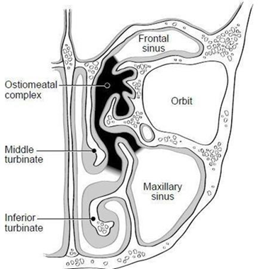
Picture 3. Complex osteomeatal piece Coronal 25
Etiopathogenesis
The etiology of antrochoanal polyps is not yet known for certain. Until now there are still many debated by for expert. However a number of factor predisposition The occurrence of polyps includes allergies, especially allergic rhinitis, chronic sinusitis, irritation, infection and nasal obstruction due to anatomical abnormalities such as septal deviation and concha hypertrophy. 13,19
On Initially, mucosal edema was found, which mostly occurred in the middle meatus area. Then the stroma will be filled with intercellular fluid so that the swollen mucosa becomes polypoid. If the process continues, the swollen mucosa will enlarge and then descend into the nasal cavity while forming stalk, so that it happened polyps. Polyps can arise In an uninfected nose, it can cause blockage resulting in sinusitis, but polyps can also arise due to chronic irritation caused by nasal and sinus infections. Polyp formation is often associated with inflammation. chronic, autonomic nervous dysfunction and genetic predisposition. 21
According to theory Bernstein, happen change mucosa nose consequence inflammation or turbulent airflow, especially in the narrow area of the ostiomeatal complex. Submucosal prolapse occurs followed by reepithelialization and formation of new glands. There is also increased sodium absorption by the epithelial cell surface resulting in retention water so that polyps are formed. Another theory says that due to vasomotor nerve imbalance, there is an increase in capillary permeability and vascular regulation disorders that result in the release of cytokines from mast cells, which will cause edema and eventually become polyps. If the process continues, the swollen mucosa will enlarge into polyps and then will descend into the nasal cavity by forming legs. 19
Occurrence of herniation antral cyst pass ostium maxillary accessory due to chronic inflammatory process due to allergies or infections. This not only causes the development of antral cysts and mucosal edema, but also causes swelling around the osteomeatal complex. Air passing through the natural ostium cannot escape during expiration due to mucosal edema and swelling in the osteomeatal complex. Based on Bernoulli's theory, higher pressure in a narrow area causes pressure to the surrounding area so that happen edema mucosa And obstruction complete on osteum experience especially during the expiration process. Continuous obstruction of the natural and accessory osteum will result in increased pressure in the Highmore antrum. This causes increased pressure in the maxillary sinus, intramural cysts can be pushed out through the accessory osteum so that they will form antrochoanal polyps. 7,10
Mac Kay And Lund share polyp in a way general become 3
Stage 1: polyps confined to the middle meatus
Stadium 2: polyp Already go out from meatus media, looks in cavity nose but does not yet fill the nasal cavity.
Stadium 3: polyp Which massive And cause obstruction total 11
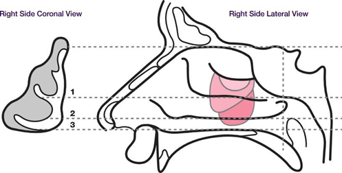
Picture 3. Polyp stage according to Mac Kay and Lund 11
Symptom Clinical
Clinical symptoms of patients with antrochoanal polyps include nasal congestion, runny nose, snoring, headache, post nasal drip , mouth breathing, epistaxis, anosmia, halitosis, dyspnea, dysphagia, dysphonia and itchy nose. Orvidas et al noted symptoms of nasal congestion in all patients with antrochoanal polyps, runny nose in about 48%, snoring in 36% and mouth breathing in 32% of patients with antrochoanal polyps. Mohd Tahir J et al studied that the most common clinical symptoms often is blockage nose (92.5%) followed with nose watery (45%), postnasal drip (35%) and snoring (22.5%). 11,20,22, 23
Macroscopic and Histopathological
In large intranasal polyps, evaluation is required. use Anterior rhinoscopy examination is very easy to perform. Macroscopically, polyps appear colored. clear grayish, shaped like fruit pear, slippery, soft And can moved. Polyps arise from lateral nasal wall And in many cases limited to meatus rice media, unilateral And on a number of case only can be seen from endoscopic examination. 10,11
Histologically, polyps consist of from fibromyxomatous tissue which is covered by The typical respiratory stromal epithelium is ciliated pseudostratified columnar epithelium and in some cases may appear as benign squamous cell metaplasia. The epithelium contains very few nerve endings and submucosal glands as well membrane basalt. If compared to with wall mucosa lateral nose adjacent, in inflammatory polyps there are eosinophils and mast cells in greater numbers. 10,11
The histopathological characteristics of antrochoanal polyps are the same as those of nonallergic maxillary sinus polyps. Antrochoanal polyps are lined by pseudostratified ciliated epithelium , with varying degrees of inflammatory cell infiltration in the stroma. The stroma is usually edematous and more vascular, consisting of loose connective tissue infiltrated mainly by plasma cells and a few eosinophils. The amount of cell infiltration inflammation far exceeds the number of eosinophil infiltrations in antrochoanal polyps when compared with nasal polyps due to allergies. 11,21
Diagnosis
Diagnosis polyp antrochoanal with enforced based on anamnesis complete, physical examination and supporting examination. From the anamnesis, the complaints that typical on polyp antrochoanal in the form of nose clogged And watery. Sometimes there is also snoring, headaches, mouth breathing, epistaxis, anosmia, halitosis, dyspnea, dysphagia, dysphonia, painful face And nose itchy. Complaint usually felt for about 1 year and even more than 3 years. 7.19
On anterior rhinoscopy examination, polyps will appear coming out of the middle meatus. This will be difficult evaluated when the polyp has filled the nasal cavity and spread to the choana and nasopharynx. will seen with rhinoscopy posterior or visible in the oropharynx behind the uvula. Polyps are often difficult to distinguish as conchae. So to differentiate, you can use a tampon given a vasoconstrictor drug, where the conchae will shrink after being given a tampon given a vasoconstrictor drug while polyps do not shrink. 8,11
Inspection Support
Inspection nasoendoscopy And CT scan is standard diagnostic antrochoanal polyp. On nasoendoscopy it appears as a smooth, pale pink or yellowish mass. On CT scan it appears as a soft tissue mass filling the maxillary antrum extending through the natural ostium or maxillary accessory ostium into the nasal cavity between the middle concha and the lateral wall of the nasal cavity, without any bone erosion or compression, then the polyp can extend posteriorly through the choana
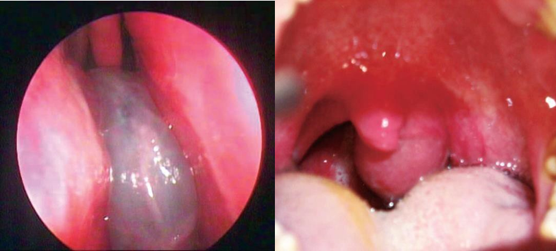
Picture 4. A. Nasoendoscopy polyp antrochoanal. 3B. Looks polyp antrochoanal behind uvula. 3
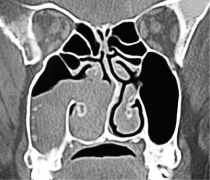
Picture . 5. A. Picture piece coronal CT scan polyp antrochoanal right 3

B. Picture axial section CT scan polyp left antrochoanal 2
Diagnosis Appeal
Diagnosis of antrochoanal polyps can be very easy if there is opacification of the maxillary antrum that is widened and a nasopharyngeal mass is visible. Other things that resemble the picture of antrochoanal polyps, need to be excluded as a differential diagnosis, including:
Hypertrophy concha inferior on Photo projection nasopharynx sometimes happen misinterpreted as a nasopharyngeal mass, but should have been easily recognized and ruled out.
Hypertrophic polyps with chronic rhinosinusitis. Etiology is associated with vasomotor instability disorders including drugs, infections, endocrine imbalance disorders and psychological factors. Allergy is the most common etiology especially in cases of bilateral polyps. Hypertrophic polyps can be unilateral or bilateral, most commonly in the nasal cavity and ethmoidal sinuses. Extension into the lumen of the sinus and nasal cavity can occur.
Juvenile nasopharyngeal angiofibroma. This tumor with high vascularization, is a benign tumor that has the potential to cause local destruction. Location in the nasopharynx or posterior nasal cavity at puberty. Symptoms include epistaxis, obstruction nose or mass in nasopharynx. Description radiology in the form of mass large soft tissue in the nasopharynx extending into the parapharyngeal area, anteriorly causing compression of the maxillary antrum on the involved side by eroding it structure bones surrounding. If need can done angiography.
Mucous retention cyst. This lesion is often in the sinus cavity, especially in the antrum. Surface fine with cyst epithelial Which containing fluid mucus, arise from the base or one wall of the sinus cavity. Does not extend into the choana or compress the sinus cavity.
Mucocele. A mucin sac. interstitial originating from rupture of glands or ducts. Usually occurs in the frontoethmoid area, rarely in the maxillary sinus. Mucocele cause enlargement And expansion sometimes very weight on the cavity and erosion or sclerosis of the cavity walls. But does not extend or pass through the choana when present in the maxillary sinus.
Malignant tumors in the nasopharynx. This tumor is present in about 1% of all malignancies. The tendency of tumors to cause bone destruction, respiratory tract disorders and invasion of the paranasal sinuses. 3,4,13
Treatment
Treatment of antrochoanal polyps can be done by surgery. The Luc-Caldwell procedure is a method that is widely known as a surgical technique for removing polyps. The Luc-Caldwell technique is a polypectomy technique that is widely used to remove polyp masses that are intra antrum. Approach through procedure Luc-Caldwell give access Which wide enough in the antrum, so that the removal of the polyp mass can be done completely. The antral part of the polyp must be taken to avoid postoperative recurrence. The side effects of the Luc-Caldwell procedure are infra-orbital nerve trauma, edema on cheek post-operative, paresthesia, disturbance growth tooth And face in children. So this surgical procedure is only performed on adult patients. 2-4
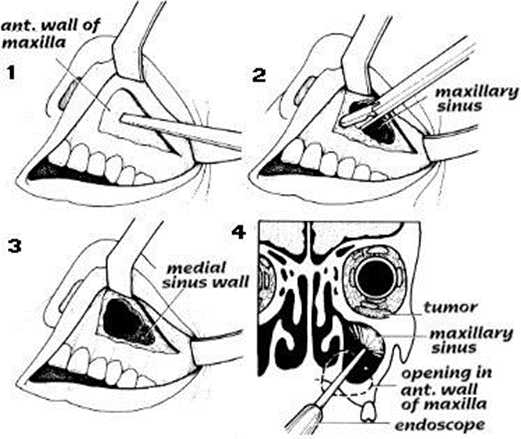
Picture 6. Procedure Luc-Caldwell. 2
At the moment surgical action with Functional Endoscopic Sinus Surgery or FESS is also quite widely chosen. The Functional Endoscopic Sinus Surgery or FESS procedure consists of resection of polyps in the more cystic intra-nasal and intra-antrum parts along with their adhesions in the maxillary sinus while maintaining the mucosa. sine normal through antrostomy meatus media. Cook et al. No find recurrence from 33 sufferer polyp antrochoanal Which Already done FESS. Oz et al. performed FESS combined with transcanine sinusoscopy or LCW on antrochoanal polyps. It was stated that there was recurrence in 3 patients after FESS and no There is recurrence on sufferer polyp antrochoanal Which done combination action FESS And Sinoscopy transcanine or LCW. Hong et al. recommended powered instrumentation using a microdebrider during FESS, as an effective technique for removing antral parts of antrochoanal polyps. No complications were found when powered instrumentation using a microdebrider and the average improvement was 96.4%. 23,
Prognosis
Antrochoanal polyps are benign lesions originating from the maxillary sinus mucosa. The prognosis is generally good, but there is a possibility of recurrence or recurrence which is very dependent on the technique of taking the antrochoanal polyp. Usually recurrence occurs because there is a part of the polyp that is not removed during surgery. The LCW technique provides good access to take the antral part of the polyp. The combination of the FESS approach with transcanine sinusoscopy or LCW is expected to be able to remove the antrochoanal polyp as a whole, both the cystic part in the sinus cavity and the polyp part in the nasal cavity so that there is no recurrence in patients. 3,21,22
Hypertrophy Konka inferior
The term turbinate hypertrophy was first introduced in 1800, which means as enlargement inferior concha and This term is still used until now. Hypertrophy is the enlargement of an organ or tissue due to the size of its cells. Which increase. On the contrary hyperplasia is enlargement Which caused by increase amount cell. Hyperplasia And hypertrophy layer mucosa And bone of the inferior concha are two factors that can explain the occurrence of inferior concha enlargement. Epidemiologically, 20% of the population with chronic nasal obstruction is caused by concha hypertrophy in a study in Europe. Causes from hypertrophy concha is rhinitis allergy And non allergy (vasomotor rhinitis) and compensation from septum deviation contralateral. Management hypertrophy Koncha can be treated with medication and surgery. Surgical therapy is performed if medication therapy fails. 5,6
Diagnostic Hypertrophy Inferior Concha
Concha hypertrophy can be assessed by anamnesis, physical examination and supporting examination. Symptoms of concha hypertrophy are nasal congestion. Assessment of the degree of this complaint can be done using the Visual Analog Scale (VAS) with a scale of 0-10. 7,16
Inspection physique with rhinoscopy front obtained hypertrophy concha.
Yanes share enlargement inferior concha become 3 namely,
concha inferior reach line Which formed between middle nasal fossa with lateral nose
enlargement concha inferior pass part from cavity rice
enlargement inferior concha has reach nasal septum 5.6
From inspection nasoendoscopy obtained enlargement concha posterior that is
concha inferior Not yet pass line koana
concha inferior has reach line koana
concha inferior has pass line koana. 5.6
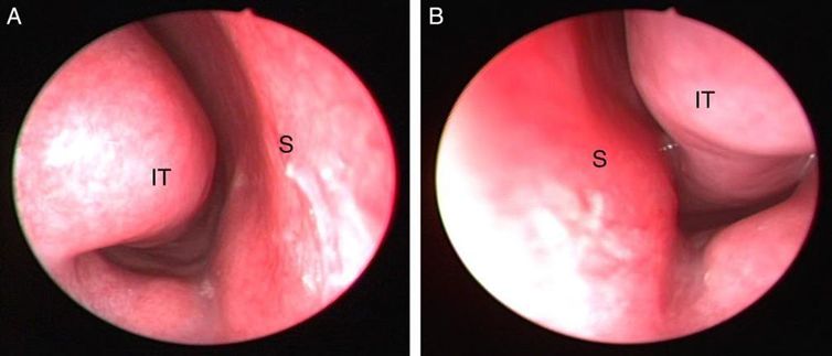
Picture 7. Endoscopy hypertrophy concha inferior 5
Cases of unilateral conchae enlargement caused by contralateral septal deviation found hypertrophy of the mucosa and bone. In general, conchae enlargement in the septum deviation is to protect the nasal cavity from dryness and crust formation due to excess air. Inferior conchae hypertrophy, part Which experience hypertrophy maximum started on side medial followed by the anterior and then posterior parts. 16,17
Bilateral inferior conchae enlargement due to allergic rhinitis is found to increase the number of goblet cells and thickening of the basement membrane. The number of blood vessels is also increased with congestion And dilation as well as obtained stromal tissue edema. Amount cell eosinophil on rhinitis allergy increase, whereas on rhinitis non allergic obtained domination gland cell mucousasinic . 16,17 Berger report There is an increase in blood vessels in vasomotor rhinitis and this phenomenon occurs in relation to the formation of new blood vessels. While in allergic rhinitis, no greater vascular density is found. 12
Inspection in lower microscope electron results biopsy concha obtained more pseudo-stratified columnar epithelium. This strengthens the statement that hyperplasia mobile proven found on hypertrophy konka. Increase wide from the mucosa medial caused lamina thickening propria that contain cell subepithelial inflammation, sinus venous And gland submucosa. The surgical target for conchae hypertrophy is the medial side and also the inferior side. Reduction in this inferior region can done Because rich will sine venous so that can reduce excessive congestion and obstruction, has few glands so that the possibility of mucosal drying does not occur and contains few arteries so that it does not increase perioperative bleeding. Lateral side of the concha must be maintained because this area is rich in glands, does not interfere with the airway, and important in guard humidity air Which inspired And maintain the normal function of the mucociliary system. In this bilateral conchae enlargement, there was no enlargement of the conchae bone. 6,16
Management Hypertrophy Konka inferior
Management of inferior concha hypertrophy can be done with drug therapy and surgery. Concha enlargement in acute cases is due to filling of the venous sinus, so that the enlargement of the concha can be reduced by administering topical decongestants. Drug therapy includes administration of antihistamines, decongestants, corticosteroids, mast cell stabilizers and immunotherapy. 11,16
Both systemic and local decongestants are effective in the treatment of nasal obstruction. Because hypertrophy concha. Usage systemic oral decongestant cause side effects such as palpitations and insomnia. Long-term use of topical decongestants can cause rhinitis medicamentosa. Corticosteroids are effective used For blockage nose, but have effect side nose easy bleeding, mucosa nose dry And formed crust. 11.16
The formation of network tie on case chronicle Which caused by chronic inflammation that no longer responds to medication after 2 months of treatment, surgery can be performed. In general, these surgical techniques can be grouped into resection and coagulation, total and partial turbinectomy, inferior turbinoplasty, submucosal turbinectomy, submucosal resection with lateral out fracture, laser, radiofrequency, electrocoagulation, argon plasma coagulation, cryotherapy And neurotectomy vidian. Objective main surgery is improve nasal breathing and maintain physiological function. There is no ideal technique, each own complications term short And term long such as bleeding and atrophic rhinitis. 11,12,16
REFERENCE
Kizil Y, Aydil U, Ceylan A, et a. Analysis of choanal polyps. Journal of Craniofacial Surgery. 2014;25(3):1082–1084.
Atighechi S, Baradaranfar MH, Karimi G, Jafari R. Antrochoanal polyp: a comparative study of endoscopic endonasal surgery alone and endoscopic endonasal plus mini-Caldwell technique. Eur Arch Otorhinolaryngol. 2009 Aug;266(8):1245- 8.
Yaman H, Yilmaz S, Karali E, et al. Evaluation and management of antrochoanal polyps. Clin Exp Otorhinolaryngol. 2010;3:110-4.
Selma et al. Antrochoanal Polyps: Analysis of 18 cases in 2006. Electronic Journal of Otorhinolaryngology. 2009.
Saulescu M, Sarafoleanu C. Surgery for Nasal Obstruction in Inferior Turbinate Hypertrophy. Romanian Journal of Rhinology. 2015; 5: 1-6.
Jourdy D. Inferior Turbinate Reduction. Operative Techniques in Otolaryngology. 2014; 25 : 160-170.
Dhingra PL, Dhingra S. Diseases of Ear, Nose, Throat, Head and Neck Surgery 6th Edition. Elsevier: India. 2013; 192-196.
Snell, RS Anatomy Clinic Based on System. Translated by Suguharto L. Jakarta: EGC. 2012.
Netter FH. Atlas of Human Anatomy. 5th ed. Philadelphia, PA: Saunders/Elsevier; 2011.
Leung RM, Walsh WE, Core RC. Sinonasal Anatomy and Physiology. Bailey's Head and Neck Surgery Fifth Edition. Philadelphia: Lippincott Williams and Wilkins. 2014; p.360.
Kenyon G. Nasal Anatomy and Analysis. Int Otolaryngol Clin. 2013; 5(1):34- 42.
Berger W E. Allergic rhinitis in children. Pediatric Drugs Journal Springer International. 2004; 6(4), p 233-250.
Frosini P, Picarella G, De Campora E. Antrochoanal polyp: analysis of 200 cases. Acta Otorhinolaryngol Ital. 2009;29:21-6.
Orv idas LJ, Beatty CW, Weaver AL. Antrochoanal polyps in children. Am J Rhinol. 2001 Sep-Oct;15(5):321-5.
Marcos Rabelo de Freitas; Rogério Pinto Giesta; Sebastião Diógenes Pinheiro; Viviane Carvalho da Silva. Antrochoanal polyp: a review of sixteen cases. Brazilian Journal of Otorhinolaryngology 2006.
Budiman BJ, Fitria H. Management of Conchae Hypertrophy. Department of Otolaryngology, Nose, Throat, Head and Neck Surgery, Faculty of Medicine, Andalas University/RSUP. Dr. M Djamil Padang. 2012; 17: 1- 10.
Saki N, Akhlagh SN, Shoar MH, Jafari NS. Efficacy of Radiofrequency Turbinoplasty for Treatment of Inferior Turbinate Hypertrophy. Iranian Journal of Otorhinolaryngology. 2011; 23(64): 87-92.
Endang M, Retno S. Nasal Polyps. In: Soepardi et al., editors. Textbook of Ear, Nose, Throat, Head & Neck Health Sciences. 7th Edition. FK UI, Jakarta, 2009:123-125.
Tahir M, et a. The Role of Powered Instrumentation in the Removal of Antrochoanal Polyps: A 10-Year Review Department of Othorhinolaryngology, Faculty of Medicine, Universiti Kebangsaan Malaysia, Kuala Lumpur. 2009; 4(2):108-14.
Peter Olsson, Prevalence of Upper Airway Symptoms and Aspects on Treatment of Nasal Polyposyis. 2009;23:114-23.
Al-mazrou KA, Bukhari M, Al-Fayez A. Characteristics of antrochoanal polyps in the pediatric age group. Annals of Thoracic Medicine 2009; 4(3):133-6.
Nikakhlagh, S Rahim F, Saki N, Mohammadi H, Yavari M. Antrochoanal Polyps: Report of 94 cases and Reviews the literature. Nigerian Journal of Medicine, Vol.21. 2012; 156-159
Oz F, Oz C, The Cagici CA, Canbolat T, Yilmazer C, Akkuzu B. Surgical approaches for antrochoanal polyps: a comparative analysis. B-ENT. 2009;4(2):93-9.
Walsh WE, Korn RC. Sino nasal anatomy, function and evaluation. In: Johnson JT, Rosen CA, editors. Bailey's head & neck surgery otolaryngology. 20th Edition5. Philadelphia: Lippincott William Wilkins; 2014:307- 19.
Hassan E, Gendeh BS, Husain S, Faizah MZ. Orbital complications secondary to acute sinusitis- a 10 year retrospective review. Eastern Journal of Medicine. 2014;19:130-6.
Comments