Dysphagia Esophageal
I Made Nudi Arthana
Dysphagia is difficulty or inability For swallow And can be classified into two main categories, namely oropharyngeal which occurs between cavity mouth And sphincter esophagus part on as well as esophageal Where happened between sphincter esophagus part on And down. Patient without symptom supra-esophageal, results test barium swallow or FEES who does not striking, And proof clinical dysphagia esophagus must referred to to gastroenterologist for barium video-esophagogram. Evaluation This must covers anatomy And esophageal motor function. Barium esophagography is relatively simple and inexpensive and can be considered as line First compared to endoscopy on dysphagia esophagus.
LITERATUR REVIEW
Anatomy Swallowing Structure
Swallow is function complex Which involving activity Which deliberate and reflex. Process This involving more from 30 nerve And muscle. Mechanism Swallowing involves the oral cavity, oropharynx, larynx, hypopharynx, and esophagus. 5
Cavity Oral
The area of the mouth where the oral phase of swallowing occurs includes the lips, teeth, gums, cheeks, oral cavity, soft palate, hard palate, and palatine tonsils. Processing food And formation bolus happen in cavity mouth. Limit Laterally and anteriorly formed by the dental arches. The superior border is formed by the palate. The tongue is located at the bottom. Posteriorly, the oral cavity changes into the oropharynx which is the superior part of the pharynx. 5

Picture 2.1 Cavity oral And pharynx. 5
Pharynx
The pharynx extends from the base of the skull to the inferior border of the cricoid cartilage at part front And limit inferior vertebra C6 in part behind. Pharynx shared into three segments, namely the nasopharynx, oropharynx, and hypopharynx. The nasopharynx is located in behind cavity nose And in on palate soft. Oropharynx extending from palate soft until bone hyoid, And hypopharynx located in behind larynx. The pharynx is tubular in shape with muscular walls consisting of longitudinal muscles on the inside surrounded by an external circular muscle layer. The external muscles consist of the superior, middle, and inferior constrictor muscles which contract play a role For narrow down wall pharynx during process swallow. Contraction in a way reflex sequentially from end superior to inferior pharynx propels food through the pharynx into the esophagus. The inferior pharyngeal constrictor muscle is the thickest of the pharyngeal constrictors and consists of two parts, the thyropharyngeus and the cricopharyngeus . Posteriorly, between the lower fibers of the thyropharyngeus and the upper fibers of the cricopharyngeus, is a small triangular area called the Killian dehiscence . This space is susceptible to diverticulum formation during abnormally uncoordinated pharyngeal peristalsis in which the peristaltic wave reaches the cricopharyngeus before it relaxes. The internal longitudinal muscles of the pharynx consist of the palatopharyngeus , stylopharyngeus , and salpingopharyngeus . These muscles shorten and widen the pharynx during swallowing by elevating the pharynx and larynx. The palatopharyngeus is the largest of the longitudinal muscles. 5
Larynx
The laryngeal framework is formed by cartilages connected by membranes and ligaments. The extrinsic and intrinsic muscles of the larynx move the framework. During swallowing, the larynx must close to prevent aspiration of food. to channel breath. Matter This achieved with lift larynx to base tongue, closure of the vocal folds, and closure of the epiglottis over the laryngeal vestibule. 5

Picture 2.2 Cartilage larynx
Esophagus
The esophagus is a muscular tube that extends from the pharynx to the stomach. It is about 23-25 cm long, starting at the lower border of the cricoid cartilage (opposite the C6 vertebra) and ending at the cardiac opening of the stomach (opposite the T11 vertebra). There are three constrictions in the esophagus, which are First is at on level sphincter cricopharyngeal namely 15 cm from tooth series. Second, it is located where the esophagus is crossed by the aortic arch and the left main bronchus, namely 23 cm from the incisors. Third, the narrowing where the esophagus penetrates the diaphragm namely 40 cm from tooth series. Wall esophagus own four layer from outside to inside namely: 4
Fibrous layer
Muscular layer (outer longitudinal layer and inner circular layer continuous with the inferior constrictor of the pharynx)
Layer submucosa (with vessels blood, nerve, mucous glands )
Internal mucosal layer (covered with a thick layer of stratified squamous epithelium with papillae on its surface)
A layer of longitudinal non-striated muscle fibers called the muscularis mucosae lies between the submucosa and mucosa. It is more prominent in the lower part of the esophagus. 4
Nerves in Swallow
Accurate harmonization is necessary between breathing and swallowing to prevent food from entering the lower respiratory tract because the pharynx serves as a common pathway for respiration and digestion. Reflex organization swallow consists of from part afferent, center, And efferent. Area receptive for reflexes include various locations in the oropharynx such as the soft palate, uvula, dorsal surface of the posterior tongue, pharyngeal surface of the epiglottis, fauces pillars, glossoepiglottic sulcus, dorsal pharyngeal wall, and pharyngoesophageal junction. Specific fluid receptors and afferent adaptive pressure receptors within the fauces pillars can trigger swallow moment respond water And touch light. Afferent primary from the mucosa oropharynx walk through nerve trigeminal, glossopharyngeal, And vagus united in tract solitary For contact synaptic with neuron order second in nucleus tractus solitarius (NTS). This reflex can be modified by lung and chest wall receptors such as vagus nerve afferents and intercostal muscles. The NTS portion is a portal afferent and also contains interneurons that carry out complex swallowing control. 4
Reports suggest that there is a swallowing center in the brain stem. The swallowing center accept fiber descendant from cortex And subcortex. Area cortical and subcortical cerebral as well as the interaction with stem brain can play an important role in the nervous regulation of the swallowing reflex. Peripheral feedback from the pharyngeal area, larynx, And esophagus modify control central swallow. Stimulation pharynx can produce increased excitability of the swallowing motor cortex. Peripheral feedback contributes to the swallowing center. 4
The trigeminal, facial, ambiguous, and hypoglossal nuclei are motor nuclei involved in swallowing. However, only a small portion of the trigeminal and facial nuclei contribute to normal swallowing. In contrast, both the ambiguous and hypoglossal nuclei activate all of their motoneurons during swallowing. 4

Figure 2.3 The process of swallowing. (A) Food (shown in green) is first chewed, mixed with water saliva For to form bolus with the proper consistency and is pressed against the hard palate by the tongue in preparation for swallowing. (B) The bolus of food is pushed into the oropharynx where the swallowing reflex is triggered. This swallowing reflex lasts about one second. (C) The epiglottis moves down to cover the airway while the striated pharyngeal muscles contract to continue moving the bolus of food past the cricopharyngeus muscle (physiological upper esophageal sphincter) and into the proximal esophagus.
Part from reflex swallow This Also in progress around One second. (D) Moment The bolus of food is propelled from the pharynx into the esophagus, contraction of the upper esophageal skeletal muscles forces the bolus through the middle and distal esophagus. The medulla controls the swallowing reflex, although the reflex can be initiated by the cerebral cortex. The lower esophageal sphincter relaxes from the beginning of the swallowing process until the bolus of food is propelled into the stomach, which can take eight to twenty seconds. 5
Anatomy Veins Internal Jugular
Veins This appear in fossa cranial posterior And go out from skull through the jugular foramen, which is located at the base of the skull. As the internal jugular vein flows into the lateral neck, it drains branches of the facial, retromandibular, and lingual veins. The course of the internal jugular vein is directed to caudal in sheath carotid, accompanied by with nerve vagus in posterior And carotid artery general in anteromedial. Veins This located appropriate in adjacent lateral And anterior to the internal carotid artery and the common carotid artery. At the junction of the neck and chest, the internal jugular vein joins the subclavian vein to form the brachiocephalic vein or innominate vein. The left internal jugular vein is slightly smaller than the right internal jugular vein. There is a valve in the vein located a few centimeters before the blood flows into the subclavian vein. 18
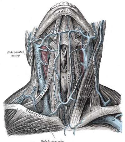
Picture 2.4 Anatomy Veins Also popular Internal 18
Physiology Swallow
Oral Phase
The oral phase of swallowing occurs in the mouth and consists of two stages: the preparatory stage and the propulsive stage. The preparatory stage begins when food or liquid is introduced into the mouth and results in the formation of a bolus. Bolus formation occurs through coordinated movements of the lips, cheeks, jaw, and tongue, as well as closure of the upper esophageal sphincter to keep the food or liquid in the mouth until the person is ready to begin swallowing. Liquids No need preparation For formation bolus, whereas Solid foods require processing to reduce particle size through chewing and softening by saliva. This process is under reflex control. through three nerve cranial. Nerve trigeminal to coordinate chewing and convey information sensory, nerve facial to coordinate movement lips and cheeks to aid in the placement and retention of food, and the hypoglossal nerve controls tongue movements. Once a bolus suitable for swallowing is formed, bolus the positioned in channel Which formed by tongue against the palate hard, And stage encouragement from phase oral started. Through elevation the tongue that wavy Which started from end And move to behind, bolus pushed backwards with a squeezing action through the fauces into the opening of the pharynx. 6
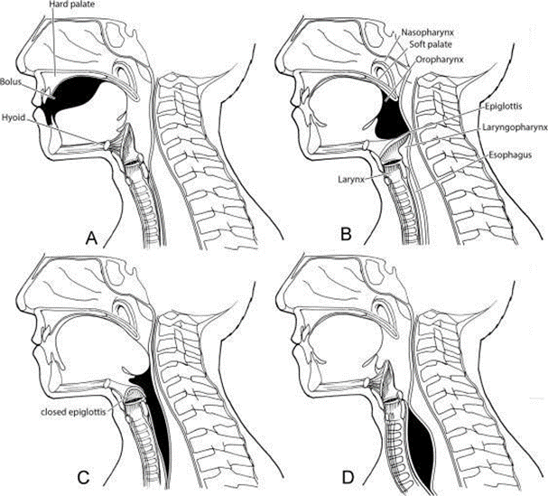
Figure 2.5 Propagation of the bolus during swallowing. (A) Wave-like elevation of the tongue propels the bolus into the oropharynx. (B) Elevation of the soft palate closes the opening of the nasal cavity as the bolus passes through the pharynx. (C) The bolus is propelled through pharynx pass epiglottis Which closed And enter to esophagus through the upper esophageal sphincter. (D) The bolus travels through the esophagus via peristaltic waves to the stomach. The soft palate and tongue are relaxed and the epiglottis is open to resume breathing. 6
Phase Pharynx
The pharyngeal phase of swallowing begins voluntarily as the bolus crosses the anterior tonsillar pillars by sensory information conveyed via the glossopharyngeal and vagus nerves. Once initiated, the remainder of this phase is reflexive and is completed in about 1 second. As the tongue propels the bolus into the pharynx, the soft palate moves upward to close the nasopharynx from the oropharynx. The palatopharyngeal folds simultaneously move toward the midline to form funnel Which guide bolus through pharynx on. Bone hyoid and larynx Then lifted in a way anterosuperior avoid from bone behind neck, which shortens and enlarges the pharynx while creating negative pressure in the hypopharynx. Propagation bolus through pharynx achieved by four mechanism following: pressure booster tongue, contraction muscle constrictor pharynx, pressure negative hypopharyngeal, and gravity. As the bolus passes through the lower pharynx, the airway is protected by elevation of the larynx beneath the base of the tongue, closure of the vocal cords, and closure of the epiglottis over the laryngeal vestibule. The pharyngeal phase ends as the bolus passes through the lower pharynx into the esophagus via the upper esophageal sphincter. 6

Picture 2.6 Muscles pharynx. (A) Side lateral from muscles constrictor pharynx. (B) Posterior side of the pharyngeal muscles cut at the pharyngeal raphe to reveal the internal longitudinal muscles of the pharynx. 6
Esophageal Phase
The esophageal phase is the reflex phase that begins when the bolus moving forward passes the upper esophageal sphincter and enters the esophagus. The upper esophageal sphincter relaxation moment phase pharynx triggered And still open until bolus pass cricopharyngeal part Which relax from constrictor pharynx inferior. By Because That, Relaxation of the cricopharyngeal muscle is essential for normal swallowing and failure to do so at the proper rate or timing can result in discoordination of the other swallowing mechanisms leading to dysphagia. Once the bolus has passed the upper esophageal sphincter, it is pushed through the esophagus into the stomach through wave peristaltic Which produce pressure positive. Secondary peristaltic waves occur in response to local distension, allowing clearance of any bolus left behind after the primary peristaltic wave. Peristaltic waves in the upper two-thirds of the esophagus move more fast than one third part lower. Matter This due to by the fact that part on esophagus contain muscle striped, whereas one third part lower non-striated. In part lower esophagus, there is segment throughout 3 cm and extends above and below the diaphragm with an average pressure that is 8 mmHg greater than the intragastric pressure. This segment forms the esophageal sphincter lower in lower contraction tonic Because strength intrinsic And extrinsic. Intrinsic forces are created by the internal circular muscle fibers of the esophagus, and extrinsic forces are created by the pressure of the diaphragm. After the bolus passes through the esophagus, powers This relax, And sphincter esophagus lower open just before the peristaltic wave carrying the bolus reaches it. 6

Picture 2.7 Wall Anterior Pharynx.
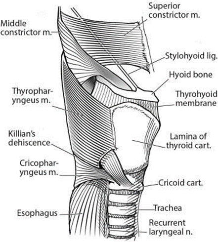
Figure 2.8 Pharyngeal Constrictor Muscles. Separation of the inferior pharyngeal constrictor into aspect thyropharyngeus And cricopharyngeus can seen on picture this. 6
Dysphagia
Oropharyngeal Pathology
Dysphagia is difficulty or inability to swallow. Oropharyngeal dysphagia is most often associated with chronic neurological conditions, especially Parkinson's disease, stroke, and dementia that are not part of physiological aging. It may also be the first symptom of neuromuscular disorders such as amyotrophic lateral sclerosis or myasthenia gravis. 5
A number of condition chronic, like health tooth Which bad, tooth false, dry mouth (xerostomia), or medication side effects may also indicate progressive oropharyngeal dysfunction. Tardive dyskinesia with choreiform tongue movements is associated with use antipsychotic term long can cause decompensation in older adults. Chronic cough associated with ACE inhibitor use may also interfere with swallowing or aspiration.
Structural abnormalities (eg, Zenker's diverticulum, cricopharyngeal bar or tumor, chronic infection with Candida or herpes virus) and extrinsic compression from cervical osteophytes or goiter can also interfere with normal swallowing. 5
Table 2.1 Reason dysphagia oropharynx 5
Chronic progressive disease (geriatric syndrome).
Stroke
Parkinson's Disease
Alzheimer And dementia other
Sarcopenia
Neuromuscular disease • Sclerosis lateral amyotrophic
Myasthenia gravity
Sclerosis multiple
Dermatomyositis/polymyositis (myopathy)
Drugs antipsychotic*.
Reason structural • Cancer head And neck
Surgery or radiation recently This for the head And cancer neck (change anatomy)
Mucositis Which induced by chemoradiation and edema (short term)
Zenker diverticulum
Osteophyte cervical
Lymphadenopathy
Goiter
Cricopharynx trunk
Reason oral • Teeth or teeth bad fake
Dry mouth (eg, xerostomia); Medications that cause dry mouth (eg, alpha and beta blockers , angiotensin converters, enzyme inhibitors , anticholinergics, antihistamines, anxiolytics, calcium channel blockers , diuretics, muscle relaxants, tricyclic antidepressants)
drugs *.
Pathology of the Esophageal
Gastroesophageal reflux disease, functional esophageal disorders, and eosinophilic esophagitis are the most common causes of esophageal dysphagia. Less common causes include medications, obstructive lesions, and esophageal motility disorders. 5
Disturbance reflux gastroesophageal (GERD) And exposure sour Which recurring causes change start from inflammation submucosa And dysmotility to erosive esophagitis and strictures. Patients with GERD may experience dysphagia even in the absence of obvious mucosal damage. Eosinophilic esophagitis is an increasingly common inflammatory condition triggered by food allergens. Chronic eosinophilic infiltration leads to progressive fibrosis of the esophageal ring and dysmotility. Like irritable bowel syndrome or dyspepsia functional, Functional esophageal disorders are thought to be caused by abnormalities in gut-brain interactions and central nervous system processing. Patients with these conditions are particularly sensitive to the amount of sour physiological. Patient Possible report dysphagia, although painful chest pain and heartburn are more common. These disorders may be the cause of many patients who report intermittent difficulty swallowing but never seek care and find no explanation even after extensive workup. Medications may cause dysphagia due to direct mucosal injury (pill esophagitis), esophageal motility disorders, or lower esophageal sphincter relaxation and reflux. Esophageal cancer, strictures, Schatzki rings , And fibrosis must under consideration on patient Which come with dysphagia. However, the prevalence of this condition is relatively low, especially in patients under 50 years of age. Esophageal motility disorders such as achalasia, distal esophageal spasm, and systemic sclerosis (scleroderma) are rare. Like opioid-induced bowel dysfunction and constipation, opioid-induced esophageal dysfunction may also occur. 5
Table 2.2 Etiology esophageal dysphagia . 6
Reason dysphagia esophageal
Disease reflux gastroesophageal And esophagitis (30% until 40%)
Esophagitis eosinophilic
Functional dysphagia
Disturbance esophagus functional (20% until 30%)
Gastritis functional
Disease reflux gastroesophageal (nonerosive)
Globe pharyngeus
Reflux Hypersensitivity Treatment (5%)
Pill esophagitis (irritation direct Which relate with ascorbate sour,
bisphosphonates, iron sulfate, drug anti-inflammatory nonsteroidal, potassium chloride, quinidine, and tetracycline)
Reflux is caused by decreased tone of the lower esophageal sphincter (associated with alcohol, anticholinergics, benzodiazepines, caffeine, calcium channel blockers, nitrates, and tricyclic antidepressants)
Condition structural or mechanical (5%)
Narrowing esophagus or peptic (caused by by erosive esophagitis )
Impaction object foreign or food (dysphagia beginning I)
Malignancy (cancer esophagus or stomach, mass mediastinum with extrinsic compression)
RingSchatzki
Disturbance motility esophagus (< 5%)
Contractility No There is
Achalasia
Spasm distal esophagus
Obstruction flow go out intersection esophagogastric
Hypercontractile (jackhammer).
Disturbance motility hypercontractile
Dysfunction esophagus consequence opioid
Infection (<5%)
Candida esophagitis
Esophagitis cytomegalovirus
Esophagitis virus herpes simplex Rheumatologic conditions (<5%)
Sclerosis systemic (scleroderma)
Diagnosis Dysphagia
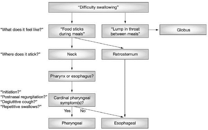
Picture 2.9 Channel diagnosis dysphagia
Channel diagnose dysphagia, There is a number of step important Which need followed, namely, identifying the presence of dysphagia, ensuring whether the patient is really experience dysphagia or only feel sensation Which similar, like globus, xerostomia (dry mouth), or odynophagia (painful swallowing). True dysphagia is difficulty swallowing that interferes with food transport, while globus is a sensation of a lump or pressure in the throat that is often unrelated to the act of swallowing and does not interfere with food transport. Then determine the Location of the Problem: Once dysphagia is confirmed, the next step is to identify whether the problem is in the esophagus or pharynx. Understanding this location is important because the type and cause of dysphagia can vary depending on which part is affected. Step furthermore differentiate abnormality structural from abnormality motor. Dysphagia can due to by problem structural, like obstruction, or motor problems involving damage to the muscles or nerves that control swallowing. Further testing, such as imaging or motor function tests, can help differentiate between the two types. 11
A number of instrument screening dysphagia Which general used is EAT-10 ( Eating Assessment Tool -10), Swallowing Disturbance Questionnaire (SDQ), Gugging Swallowing Screen (GUSS), And TIME ( Mann Assessment ofSwallowing Ability ). 6
Table 2.3 Gugging Swallowing Screen Language Indonesia (GUSS- INA)
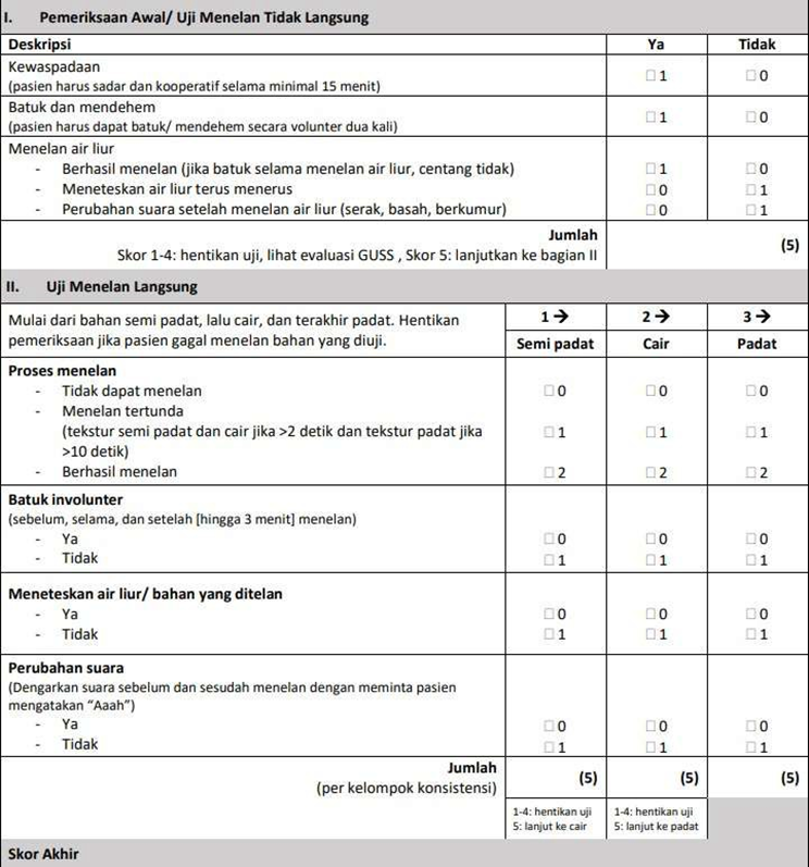
Results from dysphagia screening can help identify individuals who are at high risk for dysphagia and require further evaluation using method diagnostic Which more detailed, like FEES ( Fiber optic Endoscopic Evaluation of Swallowing ) or VFS ( Videofluoroscopic Swallowing Study ). Dysphagia screening can also be helpful in planning early interventions, such as dietary modification or swallowing rehabilitation therapy, to reduce the risk of complications associated with dysphagia. 6
Table 2.4 Questionnaire EAT-10 Adaptation Language Indonesia
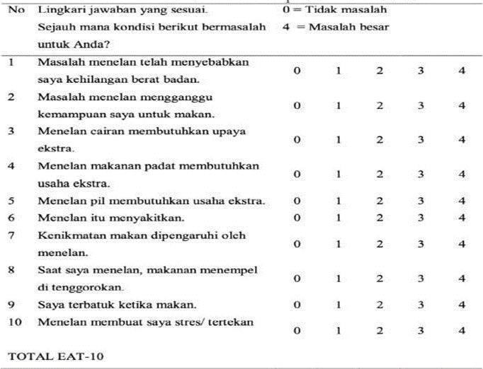
The EAT-10, is the most frequently mentioned screening instrument in the literature, and according to a number of study tool This Also capable predict aspiration risk is a questionnaire consisting of 10 questions related to dysphagia. Score Respondent For each question is 0 up to 4, Where score 0 show No There is problem And score 4 show the problem that very heavy. If score reach 3 or more show that there were swallowing problems or dysphagia in the respondents. Some of the most clinical parameters important Which displayed by EAT-10 between other heavy body, go out to eat, food solid-liquid, drugs, painful moment swallow, enjoyment, food in the throat, coughing, and stress when swallowing. 6
History Medical And Physical examination
History medical And inspection physique Which comprehensive very important in evaluating patients with dysphagia. Both can often indicate the diagnosis. 7
The medical history should include the nature of the dysphagia, the progression of the problem, and whether the problem occurs with solids, liquids, or both. Difficulty swallowing solids generally suggests an obstruction or mechanical problem such as an intermittent esophageal membrane or ring. In contrast, progressive dysphagia is more likely to be caused by a progressive stricture or malignancy. Dysphagia with liquids suggests a motility disorder such as achalasia. or sclerosis systemic. If nature intermittent, so Possible caused by spasm esophagus. A number of matter Which need asked on history including: 7
Chronology beginning
Duration And development of symptoms
Problem with initiation swallow or after a number of swallowing seconds
Symptom especially in neck or chest
Happen with food congested, fluid, or both of them
Change diet Which done Alone related with consistency food; preference for liquids or semisolids
Eat slowly
Cleaning throat during or after Eat
Cough, choked, or regurgitation postnasal Which related
History infection chest recurrence
Decreased function
Neurological deficit
Decrease heavy body
Feeling Embarrassed
Isolation social, frustration, or depression
Signs physique can includes: 7
Lost tooth or disease gingiva
Closing lips Which abnormal
Change mucosa local
Change neurological
Difficulty initiating a swallow is more typical of oropharyngeal dysphagia and esophageal dysphagia usually occurs within seconds of swallowing. Clinicians need to differentiate between both of them Because often time is process pathological different . 7
Symptom dysphagia oropharygeal: 7
Delay initiation swallow oropharyngeal
Cough deglutition
Aspiration nasal during swallowing
Need swallow repetitive For clean nasal secretion
Sensation globe
Symptom dysphagia esophagus: 7
Symptom appear a number of second after initiation swallow
Regurgitation food Which No digested
Dysphagia feel more in in chest
Although dysphagia oropharyngeal often seen on population geriatrics, smoking history or use alcohol, with or without decline heavy body Unwanted findings suggest the possibility of underlying malignancy. Regurgitation of undigested food, hoarseness, halitosis, a feeling of fullness in the throat, and a history of aspiration may indicate Zenker's diverticulum which is believed to result from weakness of the cricopharyngeal muscle. 7
Inspection Support
Oropharyngeal Dysphagia
In patients with clinical evidence of oropharyngeal dysphagia, the initial test is a clinical swallowing evaluation performed by a speech pathologist or nurse. However, test This own limitations And Possible No detect more from 50% of patients with aspiration. Early detection of oropharyngeal dysphagia using screening tools can reduce complications, especially in the older population. Several screening evaluation tools are available, but they have poor predictive value for aspiration.
After evaluation swallow clinical, two test instrumental often used to assess oropharyngeal dysphagia. Videofluoroscopic swallow study (VFSS) allows direct visualization of bolus passage by evaluating swallowing physiology, swallowing efficiency, and aspiration risk through a two-dimensional view. Although this study has significant diagnostic value, scientific tools need to be developed to improve reliability and reduce inter-reader variability, which is a limitation of this study. Fiberoptic endoscopic evaluation of swallowing (FEES) is performed by inserting an endoscope through nose For observe nasopharynx, oropharynx, And larynx to allow direct visualization and detailed analysis of the swallowing structures and their function during the consumption of foods of varying consistencies. 7
The choice of test is determined by the characteristics of the patient. To perform VFSS, the patient must be able to sit upright and mobilize to the radiology department. In contrast, FEES can be performed in bed. The results of a recent systematic study showed that FEES is more sensitive in detecting penetration, residue, and aspiration than VFSS. However, both are the gold standard for assessing dysphagia related to Parkinson's disease. In addition, high-resolution manometry can complement other diagnostic strategies to assess upper esophageal sphincter pathology, especially in patients with globus sensation. 7
Patients with signs of malignancy, such as weight loss or bleeding should undergo a head and neck evaluation and laryngoscopy. from pharynx And larynx. Attention special must given on oropharynx and possible Zenker's diverticulum. Esophagography usually diagnoses Zenker's diverticulum. In addition, computed tomography of the head and neck may be considered to rule out malignancy. 7
Dysphagia Esophageal
Patient without symptom supra-esophageal, results test barium swallow or FEES who does not striking, And proof clinical dysphagia esophagus must referred to to gastroenterologist for barium video-esophagogram. Evaluation This must covers anatomy And esophageal motor function. Barium esophagography is relatively simple and inexpensive and can be considered as line First compared to endoscopy on dysphagia esophagus.
Endoscopy is associated with risks such as aspiration, especially in elderly patients . Barium video-esophagography is also useful in the diagnosis of achalasia and can help objectively document treatment outcomes and detect recurrence early. Barium video-esophagography should also be considered as a test beginning when cancer ring esophagus, stricture, or esophagitis reflux weight is suspected with a sensitivity of 90% to 95%. 7
Choice test beginning must based on on characteristics patient And shared decision making. The combined approach of barium esophagography and endoscopy can done. Lots gastroenterologist recommend endoscopy as initial test because most patients who undergo esophagography eventually require endoscopy. Endoscopic evaluation provides additional benefits such as therapeutic interventions such as dilation and biopsy. 7 High-resolution esophageal manometry is preferred over conventional manometry. It is useful in the diagnosis of achalasia, diffuse esophageal spasm, jackhammer esophagus , and esophagogastric outflow obstruction. 7
The use of Transnasal Esophagoscopy (TNE) is increasing worldwide in the field of otolaryngology as an investigative and interventional tool in patients with globus reflux and dysphagia. This procedure has been widely validated since its first use. from esophagus oral to nasal intubation use scope fiber optics flexible esophagoscopy in the 1990s. Jonathan Aviv, an American head and neck surgeon, developed the technique and demonstrated that it could be performed on an outpatient basis, without sedation. Since then, TNE has been used to perform biopsies, balloon dilation of the throat, vocal cord medialization, and secondary tracheoesophageal puncture. The main advantage of TNE compared to esophagoscopy, with oral intubation, or rigid esophagoscopy, under general anesthesia, is that the patient does not require sedation or anesthetic total. Matter This, on its turn, produce reduce time take care hospital stay and shorter procedure time. 14
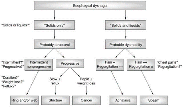
Picture 2.10 Channel diagnosis on dysphagia esophageal
If esophageal dysphagia is suspected, the next step is to determine whether the dysphagia is caused by a structural or motor disorder. Dysphagia that is worse with solid foods usually indicates a structural disorder, such as a stricture or tumor, that is blocking the passage of food. If dysphagia happen with food liquid And congested, This often leading to problem motor, like achalasia or spasm esophagus, in where muscles or the nerves that control swallowing may not function properly. Then based on progression, if dysphagia is progressive, where symptoms get worse over time, it is more often associated with structural abnormalities, like enlargement tumor. Dysphagia Which appear And is lost more often occurs in motor disorders, where the esophageal muscles may function well on some occasions but not consistently. Odynophagia or pain when swallowing may occur which is due to inflammation or infection of the esophagus. And Can happen Good on abnormality structural and motor, although it occurs more frequently in certain conditions such as esophagitis. 11
Veins Jugular Phlebectasis
Definition Veins Jugular Phlebectasis
Jugular vein phlebectomy means dilatation of the jugular vein. This condition is commonly observed in children or young adults. Jugular vein phlebectomy internal usually appear as swelling Which can happen neck, consistency soft, usually grow bigger with maneuver Which increase pressure intrathoracic like maneuver Valsalva, sneeze, or cough, And usually unilateral. 8
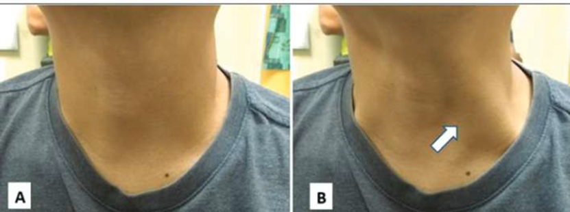
Picture 2.11 Veins jugular internal (arrow) A) moment Rest And B) enlarged during the Valsalva maneuver. 16
Epidemiology Veins Jugular Internal Phlebectasis
Internal jugular phlebectasis is a rare disorder that can occur unilaterally. or bilateral, And often diagnosed during time children. Thus, the occurrence of internal jugular phlebectasis in adults is compared with the emergence on time children very much seldom occurred. 12 Several cases have been reported to have experienced internal jugular phlebectasia appearing in age mature; However, swelling neck seen clear since born on both cases said. 12 The incidence of this condition is rare and only about 100 cases of phlebectasis have been previously reported. The jugular veins involved include the internal jugular vein as the most common, followed by the external jugular vein and the anterior jugular vein. 8
Etiology Veins Jugular Internal Phlebectasis
Scarcity condition This so that Not yet There is possibility etiology Which can be found. However, hypothesized that lack of elasticity main wall veins may be the reason that explains most of the congenital cases. In addition, increased pressure in the internal jugular vein is another hypothesis that can cause phlebectasis. 8
A number of writer has to hypothesize that injury direct previously on the neck or procedure medical like catheterization vein central, ventilation pressure positive, neck surgery, or tumor. Histopathological changes in cases treated with resection surgery covering depletion layer muscle And elastic. However, most of the case No show change significant compared to with normal venous wall. Several authors have found this abnormality in patients with neurofibromatosis type 1 and Ehlers-Danlos syndrome. 8
Pathophysiology Veins Jugular Internal Phlebectasis Causes Dysphagia
The term phlebectasis differs from aneurysm. Phlebectasis indicates a homogeneous fusiform dilatation along the vein, whereas an aneurysm is characterized by localized ectasia. Phlebectasis also differs from varicose veins in that it lacks tortuosity. Throughout vein jugular internal there is structures important in swallowing such as nerves and anatomical structures. Voice changes, paralysis of the vocal cords voice, and/or dysphagia is a number of symptom Which can observed. The proximity of the jugular vein to the vagus nerve and other lower cranial nerves is a cause from symptoms This. Besides That, dysphagia can due to by obstruction of the canal. Mass big can influence channel Which make patient experiencing dysphagia. 9
This condition has also been reported in a case involving a boy. aged 7 month with phlebectasis vein jugular internal right Which large mass causing dysphagia. The mass was removed via a cervico-thoracic approach. with repair symptom post operation. Opposite with Which Hopefully, the ectasia is not fragile and is covered with a pseudocapsule of fibrofatty tissue which makes removal easier. 3
Phlebectasis vein jugular internal usually appear in side right with right and left ratio 5.2:1.0. 12 Internal jugular vein phlebectomy is more frequently seen on the lateral side. right Because valve vein almost No Once seen on vein right brachiocephalic vein. 13 LaMonte et al hypothesized that phlebectasis is mostly right-sided because the right innominate vein is in contact with the pleura. apical right. 13 By Because That, every improvement pressure intrathoracic can be directly channeled into the right internal jugular vein. 12
Inspection Support
Inspection Radiology
The pathology detected or the diagnosis made is related to the symptoms. Seen There is mass Which stand out in side neck moment do Valsalva maneuver. On physical examination, the mass is not solid and easily compressed. Supporting examinations that can be done with color Doppler USG (DUS), Computerized tomography (CT) angiography are performed to confirm the exact diagnosis. 26
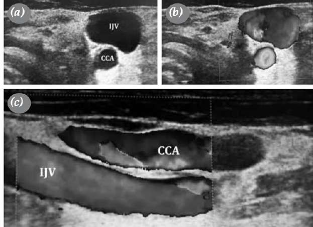
Picture 2.12 (a) Picture USG show phlebectasis vein jugular internal.
(b) USG Doppler colored show phlebectasis vein jugular internal. (c)
USG Doppler colored show parallelism artery carotid Communist And internal jugular vein on the longitudinal axis. 26

Figure 2.13 Computed tomography angiography image showing phlebectasia . vein jugular internal left. (a) slice coronal, (b) sagittal, And (c) transversal. 26

Figure 2.13 Three-dimensional reconstruction of a computed tomography angiography scan. (a) Looks front (arrow show phlebectasis vein left internal jugular), (b) left lateral view, (c) posterior view. 26
Inspection Transnasal Esophagoscopy (TNE)
Transnasal Esophagoscopy (TNE) is a widely used tool in diagnosis And treatment patient Which own complaint in in head And neck. This is because this additional investigative examination provides the advantage of visualize in on level muscle cricopharynx If compared to with esophagogastroduodenoscopy (EGD). 19
Transnasal esophagoscopy is performed using topical anesthesia without sedation. Anesthesia topical given on cavity nose with use cotton lidocaine 2% to get the effect of vasoconstriction and reduce pain, then two sprays of xylocaine 10% sprayed on the oropharynx and hypopharynx to reduce reflex gag reflex and reduce pain. Flexible esophagoscope with a 5.8 mm diameter scope using the Olympus Evis Excera II camera system. This tool has a water flushing system and air suction, as well as suction through the available working channel. 20
Table 2.5 Indication relatively Transnasal Esophagoscopy 21 Dysphagia
Odynophagia
Swallow food Which contains caustic
Symptom reflux gastroesophageal Which Already in progress long (GERD/regurgitation) Symptoms esophagus Which settle down although Already get treatment proper reflux
Evaluation foreign object
Evaluation esophagus consequence varicose veins esophagus Anorexia
Therapy
Injection Installation toxin botulinum stricture dilation Ensoscopic ablation
Therapy Placement capsule telemetry pH Wireless Tracheoesophageal puncture

Figure 2.14 Overview of TNE examination results a) Shows cardiac insufficiency. (b) shows hiatal hernia. (c) Lymphoid follicles. highlighting mild mucosal inflammation. (d) Visible reflux, chromoendoscopy (bottom image) shows white lymphoid tissue and dark green dilated blood vessels highlighting mild mucosal inflammation. (e) Peptic esophagitis, showing some erosive lesions on lower esophagus (f) Contractions tertiary, show contraction esophagus Which No coordinated due to reflux. (g) Upper esophageal sphincter is inadequate with a permanent gap.
(h) Mucosa stomach ectopic show island small mucosa stomach ectopic below the upper esophageal sphincter. 22
TREATMENT
Veins Jugular Internal Phlebectasis
Lesion tend For handled in a way conservative For avoid air embolism, venous thrombosis, and injury to important structures in the neck during dissection. Several veins and plexuses that drain into the internal jugular vein are not only close with its formation in base skull but Also during his journey in neck. Communication Which Already There is This help in flow flow veins to the opposite internal jugular veins as many of these are likely to have been opened preoperatively due to turbulence in phlebectasis.3
Ligation unilateral vein can cause edema cerebral And increased intracranial pressure, especially if the vein is the dominant vein and the vein is contralateral No capable to balance flow go out. Although seldom, problem This may occur during surgery or transiently in the immediate postoperative period. Transient elevation of blood pressure in patients may trigger the Cushing reflex . No long-term complications have been observed in the literature. Removal has been the most common mode of management. 3
Dysphagia Management
Modification diet
Diet modification can be done by eliminating foods with certain consistencies or sizes that can cause dysphagia. Modification of bolus consistency should be done given as a last resort due to changes diet often No comfortable for patient. If therapy other No To prevent aspiration or improve swallowing effectiveness, certain consistencies must be eliminated from the patient's diet. In addition, patients are also recommended to eat food in smaller sizes and with a smaller consistency. gentle (especially on patient with dysphagia oropharyngeal). Patient Which not possible to swallow or the swallowing process has a high risk of aspiration, a nasogastric tube can be installed to maintain enteral nutrition (for example in patients with stroke). To help patients with dysphagia, food is usually modified into certain textures, including 23,24 :
Pureed food : Food processed until it has a soft texture like porridge or mashed potatoes, suitable for patients who have difficulty swallowing solid foods.
Soft food : The texture is denser than puree but still easy to chew and swallow, such as pudding or steamed fish .
Fluid thick ( thickened liquids ) : Fluid Which modified consistency so that it does not easily enter the respiratory tract, for example by using a special thickener for water, juice or milk.
Exercise Postural And Maneuver Swallow
Swallowing rehabilitation is generally performed on stable dysphagia patients and on patient dysphagia with disease chronic progressive (for example disease Parkinson's). Rehabilitation swallow This aiming For strengthen muscles jaw, lips, and tongue. In addition, patients are also asked to always eat in an upright position. Some postural exercises that can be done include chin tuck, head rotation to the affected side, a combination of head rotation and chin tuck, side-lying posture, and head tilt 2 . The chin-tuck maneuver helps push the food bolus towards the pharynx and esophagus, compensates for the delay in glottis closure, and reduces the risk of aspiration during the swallowing process. 23,24
There are 4 swallowing maneuvers that can be performed by patients with dysphagia, namely supraglottic swallow, super-supraglottic swallow, effortful swallow , and Mendelsohn maneuver. The purpose of the Mendelsohn maneuver is to increase the duration and elevation of the larynx, thereby increasing the opening of the cricopharyngeal. 25
Diagnosis Appeal
Diagnosis appeal covers laryngocele, hygroma cystic, cyst branchial, and superior mediastinal mass. Chest radiography can exclude the diagnosis of laryngocele or mediastinal tumor due to the absence of air or a wide mediastinum on the film. The diagnosis is confirmed by ultrasonography combined with Doppler imaging or contrast-enhanced spiral computed tomography (CT). Color Doppler as an inexpensive technique is the diagnostic modality of choice, especially in developing countries. 8
REFERENCE
Roberts H, Lambert K, Walton K. The Prevalence of Dysphagia in Individuals Living in Residential Aged Care Facilities: A Systematic Review and Meta- Analysis. Healthcare. 2024 Mar 13;12(6):649.
Adkins C, Takakura W, Spiegel BMR, Lu M, Vera-Llonch M, Williams J, et al. Prevalence and Characteristics of Dysphagia Based on a Population Based Surveys. Clinical Gastroenterology and Hepatology. 2020 Aug;18(9):1970-1979.e2.
Cook IJ. Diagnostic Evaluation of Dysphagia. Nature Clinical Practice. 2008;5(7):393-403.
Sundaram J, Menon P, Thingnum SKS, Rao KLN. Dysphagia because of unilateral internal jugular veins phlebectasia in an infant. J Pediatr Surg. 2016 Jul;51(7):1216– 9.
Rathod P V., Vidhyadharan S, Iyer S. Anatomy of Swallowing. In: Dysphagia Management in Head and Neck Cancer. Singapore: Springer Singapore; 2018. p. 3–26.
Wilkinson JM, Codipilly DC, Wilfahrt RP. Dysphagia: Evaluation and Collaborative Management. Am Fam Physician. 2021 Jan 15;103(2):97–106.
Hennessy M, Goldenberg D. Surgical anatomy and physiology of swallowing. Oper Tech Otolayngol Head Neck Surg. 2016 Jun;27(2):60–6.
Azeri SA, Cannula AK, Kshirsagar RK. Dysphagia. 2024.
Women's A, Goyal A, Kumar A. Phlebectasia of Internal Jugular Vein- A Rare Differential Case of Neck Swelling With Review of Literature. Iran J Otorhinolaryngol. 2019 Jul;31(105):239–42.
Figueroa-Sanchez JA, Ferrigno AS, Benvenutti-Regato M, Caro-Osorio E, Martinez HR. Internal jugular phlebectasia: A systematic review. Heaven Neurol Int. 2019 Jun 19;10:106.
Basbug HS. A rare internal jugular veins phlebectasia: Reviews of the literature. Turk Gogus Kalp Dama. 31 Oct 2016;24(4):759–62
Malik V, Kumari A, Murthy T. Unusual case of focal neck swelling: Phlebectasia of internal jugular vein with intracranial extension. Int J App Basic Med Res. 2015;5(1):58
Rocke J, Ahmed S. Transnasal Esophagoscopy—Our Experience. Int Arch Otorhinolaryngol. January 2019;23(01):007–11.
Rivard AB, Kortz MW, Burns B. Anatomy, Head and Neck: Internal Jugular Veins. [Updated 2023 Jul 24]. In: StatPearls [Internet]. Treasure Island (FL): StatPearls Publishing; 2024 Jan-. Available from: https:// www.ncbi.nlm.nih.gov/books/NBK513258/
Haney JC, Shortell CK, McCann RL, et al. Congenital jugular vein phlebectasia: a case report and review of the literature. Ann Vasc Surg 2008; 22(5): 681–683,
Jian Hong L, Snow J, Tingze H. Surgical treatment of jugular veins phlebectasia in children. Am J Surg 2006; 192(3): 286–290
Standring S, et al. Gray's Anatomy The Anatomical Basis Of Clinical Practice 41st ed . Elsevier Limited. 2016.
Walton J, Silva P. Physiology of swallowing. Surgery (Oxford). 2021;39(9):563- 568.
Rocke J, Ahmed S. Transnasal Esophagoscopy—Our Experience. Int Arch Otorhinolaryngol. January 2019;23(01):007–11.
Prime RF. Transnasal Esophagoscopy Examination in Outpatient Unit, Department of Otorhinolaryngology Head and Neck Surgery, Dr. Soetomo Hospital, Surabaya. Folia Medica Indonesia. April 3, 2020;56(1):75.
Belafsky PC, Postma GN, Daniel E, Koufman J.A. Transnasal Esophagoscopy. Otolaryngol Neck Surg. December 2001;125(6):588–9.
Horvath L, Fostiropoulos K, Burri E, Kraft M. Value of Transnasal Esophagoscopy in the Workout of Laryngo-Pharyngeal Reflux. JCM. 20 July 2021;10(14):3188.
Crowson MG, barrel BC, Lee HJ, Song Y, Miso S, Jones HN, Cohen S. Vocal Fold Paralysis/Paresis US a Markers for Poor Swallowing Outcomes After Thoracic Surgical Procedures. Dysphagia. 2019;34(6):904–915.
Verma A, Hadaya J, Tran Z, Dobaria V, Madrigal J, Xia Y, et al. Incidence and Outcomes of Laryngeal Complications Following Adult Cardiac Surgery: A National Analysis. Dysphagia. 2022;37:1142–115.
Ekberg O. Dysphagia Diagnosis and Treatment 2nd Edition. Springer Nature;
,2019
Asouhidou I, Natsis K, Asteri T, Sountoulides P, Vlasis K, Tsikaras P. Anatomical variation of left internal jugular vein: clinical significance for an anesthesiologist. European Journal of Anesthesiology. April 2008;25(4):314–8
Comments