OBJECT FOREIGN FLAKES BONE IN ESOPHAGUS THE MIGRATED WITH COMPLICATION OF ESOPHAGEAL ABSCESS
I Made Nudi Arthana
ESOPHAGEAL ABSCESS caused by Object foreign in esophagus is object or food Which stopped in in the esophagus And No can enter to in stomach. Swallowed object foreign occurs frequently in people in Southeast Asia, with the type of foreign object swallowed in adults being swallowed like fish bones which occurred in 60% of all cases, followed by chicken bones 16%. 12 In Indonesia, based on research from January 2014 to December 2016 in the Section ENT-KL General Hospital Prof. Dr. R.D. Kandou Manado incident Esophageal foreign bodies aged 0-10 years around 32.1% of the total 56 cases and aged 51 years and above with an incidence of 19.6% of cases. 22
LITERATURE REVIEW
Anatomy of the Esophagus
Esophagus is a tube neuromuscular Which connect hypopharynx with stomach. Esophagus on person mature own long around 23-25 cm and extends from the inferior border of the cricoid cartilage (C6) to the cardiac orifice of the stomach (T10). The esophagus is located behind the trachea and heart, passing through the mediastinum and the hiatus, an opening in the diaphragm, on its way down from cavity chest to cavity abdomen. Esophagus No has a serous layer; the tissue around the esophagus is called adventitia. 1,2

Picture 1. Anatomy Esophagus 1,2,3
The proximal or upper part of the esophagus is called the esophageal introitus which is located at the level of the lower border of the cricoid cartilage and the final end of the esophagus is at the diaphragm. The shape of the esophageal introitus is not round, its transverse diameter is 23 mm and its antero-posterior diameter is 17 mm. 3,4 On the ventral side of the esophagus are the trachea, left bronchus, pericardium, and diaphragm. On the dorsal side of the esophagus are the ventral plain of the vertebral column, right intercostal artery, thoracic duct, and hemiazygos vein. 4,5
Esophagus shared become three segment anatomy, that is cervical, thoracic, and abdominal. 1,2,3 The cervical segment extends from the lower end of the pharynx (at the level of the 6th vertebra or the lower border of the cricoid cartilage) and extends to the inlet thorax ( suprasternal notch ); 18 cm from incisor. Segmen thoracic extends from the thoracic inlet to the level of the tracheal bifurcation; 18 until 23 cm. Part middle thorax elongated from branching trachea midway to the gastroesophageal junction ; 24 to 32 cm. Thoracic segment lower elongated from mid between branching trachea and gastroesophageal junction , including abdominal esophagus ; 32 until 40 cm. 3 Esophagus own four constriction on track vertical, that is first narrowing of the upper esophageal sphincter in the cricopharyngeal region Which due to by emphasis muscle cricoid And cricoid cartilage at the level of vertebra VI or 14-16 cm from the upper incisors (Figure 2A), The second esophageal narrowing occurs due to the crossing of the aortic arch at the level of the vertebrae. thoracic IV or 23 cm from incisor on, Where in area This it seems there is pulsation aorta (Picture 2B), constriction third due to by compression of the left bronchus at the level of the V thoracic vertebra or 27 cm from the upper incisor (Picture 2C), And constriction fourth in area sphincter esophagus low part that is on time esophagus penetrate diaphragm (hiatus diaphragm) as high as vertebra Thoracic X or 38-41 cm from upper incisor (Picture 2D). 1,2,5 In area cricopharyngeus is area Which most just had time in all channels digest so that in area This most often stuck object foreign as well as at risk the occurrence perforation esophagus due to instrument.1,3,4

Picture 2. A Sphincter esophagus part on. B. Narrowing in aortic arch crossing. C. Narrowing of the left bronchus compression. D. Lower esophageal sphincter. 1,2,5
The esophageal wall consists of four layers, namely the mucosa, submucosa, muscularis and the outer layer which consists of sparse connective tissue. Different with area other on channel digestion, esophagus No has a serous layer. This causes the esophagus to be more sensitive to mechanical trauma. 4.6 The mucosa is formed from stratified squamous epithelium that changes into columnar epithelium in the area bordering the esophagus and stomach which is called the "Z" line. The submucosal layer consists of elastic and collagen layers and contains secretory cells that produce mucus. The upper muscularis layer of the esophagus is skeletal muscle consisting of circular muscle on the inside and a layer of longitudinal muscle on the outside. While the muscles in the lower half are smooth muscles, the part of which consists of a mixture of skeletal muscle and smooth muscle. 4.6
The main innervation of the esophagus is carried by sympathetic and parasympathetic fibers of the autonomic nervous system. The parasympathetic fibers are carried by nerve vagus. Distribution blood esophagus follow pattern segmental, top supplied by branches artery thyroiditis inferior And subclavian. Middle part supplied by branches segmental aorta And artery bronchial, while the subdiaphragmatic part is supplied by the left gastric artery and the inferior phrenic artery. 6
Object Foreign Esophagus
Definition And Classification
Esophageal foreign bodies are objects or foods that are stuck in the esophagus and cannot enter the stomach. In general, foreign bodies can be divided into organic and inorganic foreign bodies. Example object foreign organic is piece food, flakes bones, grains, and teeth. While examples of inorganic foreign objects are money. metal, pin, battery, And rock. Besides two Category in on, foreign object can shared based on its characteristics, like sharp, blunt, and corrosive. Battery button is type object foreign Which is corrosive and must be removed immediately because it can cause esophageal perforation within hours. 2.5Type object foreign varies on every age group. Coin or coins are a type of foreign object found in the population children. Whereas on population mature, flakes Bones are the most common finding. Foreign bodies can become lodged anywhere along the esophageal lumen, but they are more likely to become lodged in the area constriction esophagus, Good Which experience and also Pathological. The most common location for foreign bodies to get stuck is in the cervical esophagus, at the level of the cricopharyngeal muscle. 5,6
Epidemiology
Swallowed object foreign complaint Which often happen on resident in Southeast Asia. According to Tong Lim Lu et al, the types of foreign objects swallowed in mature, often found swallowed bone fish Which happen by 60% of all cases, followed by chicken bones at 16%. This is due to the nature of fish bones which are slender, less rigid and almost invisible when mixed with rice. 12 Research on the ingestion of object foreign on age mature Also has observed on study in Department Gastroenterology, House Sick Tianjin Medical University General involving 1,131 patients with 90.16% case found object foreign in channel digest. Predictor The main criterion for the presence of a foreign body is presentation less than 24 hours. Type object foreign Which found is jujube pits (36.72%) and fish bones (22.00%), and more than 80% of foreign objects were found in esophagus. 10 In Indonesia, based on research period January 2014 to December 2016 in Part ENT-KL General Hospital Prof. Dr. RD Kandou Manado, the incidence of esophageal foreign objects in children aged 0-10 years is around 32.1%. of the total 56 case And age 51 year to the top with incident 19.6% cases. 22 Studies show that most food impactions occur in adults over the age of forty, often accompanied by disturbance motility esophagus or abnormality structural in the esophagus. In children, consumption of foreign objects often occurs due to curiosity. experience, especially between age six month until three years. Although most swallowed foreign objects will pass out naturally experience, around 10-20% need intervention endoscopy, and less from 1% need operation. 9 Study Which done in Hi-Tech Medical College & Hospital find that children aged between 2-10 years are the most affected by swallowed object foreign. Majority case in study this is caused by swallowing coins, followed by thorns fish, bones meat, And safety pin. 8
Foreign body ingestion mostly (> 75%) occurs in adults. after age decade fourth life And majority have esophageal motility disorders and/or esophageal pathology. 8,9 The types of objects that are most often swallowed by adults come from food, usually in the form of pieces of meat and bones, and dentures. 11 According to Magalhães-Costa P et al., the most common foreign objects swallowed on person mature including bone fish (9-45%), bones (8-40%), And tooth false (4-18%). Object foreign Which sharp is Which most feared in stomach And intestines duodenum, Because type object this is associated with level perforation until 35%. Object Which more big from 2 cm or more long from 5 cm will experience difficulty to cross pylorus, pass ligament Treitz, And ileum. 9 On child – children in about 80% of cases swallow non-food foreign objects such as coins, buttons, small toys, and marbles. 9
Pathophysiology Object Esophageal Abnormalities
Esophageal foreign bodies are all objects, whether swallowed intentionally or not, that can cause esophageal obstruction and injury. Types of foreign objects that are often swallowed by people mature originate from food, usually in the form of piece meat, bones, and dentures. Small toys and coins are often found in children. Foreign objects, whether sharp or blunt, that are small in size can easily pass through the upper digestive tract to the stomach, except for larger foreign objects that are somewhat difficult to pass, so assistance is needed. endoscopy For take it out. Sometimes Although small but relatively made of heavy metal, like coins are rather difficult to pass through the stomach. 3,5,10
Object foreign ingestion can stuck Where just throughout esophageal level, but is most common in physiological narrowing or pathological. Place most frequently stuck is in esophagus part cervical appropriate in lower constriction cricopharyngeal. Jackson (1950) suspected that weakness in the upper cervical peristaltic muscle structure was the main cause of foreign object entrapment. It is believed that the area the is transition from muscle striped become muscle plain. If happen incarceration on esophagus thoracic or on place further down, it is necessary to think about the presence of underlying esophageal disorders. On the other hand, if there is food impaction in the lower esophagus middle or lower, matter This Possible due to by existence lesi
esophageal or presence of motor disorders. Various conditions that can underlie the occurrence of esophageal foreign bodies include reflux esophagitis which accompanied by stricture, stenosis post operation, web, Schatzki rings , hiatal hernia , cardioachalasia , carcinoma, diverticula, and motor disorders. 7The esophagus is a fibromuscular tube that begins at about the sixth cervical vertebra, located behind the cricoid cartilage. This across mediastinum from on to lower. Esophagus low part to form arch in around aorta thorax, passes behind the heart, and after the tracheal branches, passes posterior to the pulmonary artery right, atrium left, And bronchus main left. Then, the esophagus penetrate diaphragm in around vertebra thorax tenth and ends in the stomach with a length of about 25 cm. On its way, the esophagus own four point Which often experience blockage or impaction: 1) in behind bone vulnerable cricoid on muscle cricoid, 2) as it crosses in front of the aortic arch, 3) at the level of the left main bronchus, and 4) at the esophageal hiatus as it penetrates the diaphragm. Approximately 75% from object foreign Which swallowed tend happen on level muscle cricopharynx

Figure 3. Most common locations of esophageal obstruction/impaction
Swallowing object foreign on channel digestion part on is situation Which often happen And influenced by various Risk factors for foreign body ingestion in adults include the elderly population, patients with behavioral disorders, and those Which own factor risk physique like stenosis peptic ulcer or neoplastic, esophageal motor disorders, or esophageal diverticula. The act of ingesting a foreign body can occur accidentally or intentionally, and the nature and location of the object can determine urgency extraction endoscopy. Object Which more small from 2 cm can usually pass through the digestive tract without complications, but extraction may need to be considered if the object does not pass through the pylorus after 3-4 weeks or if the patient is symptomatic. Sharp objects such as needles, nails, toothpicks, and pins should be removed before they reach the stomach because of the high risk of causing perforation. 13
Pathophysiology of Foreign Bodies in the Esophageal Migration Swallowed bone fish, Which own characteristic thin, linear, and sharp, can pose a risk of penetration of the esophageal wall or extraluminal migration to surrounding structures in the neck, Which on Finally can cause serious complications. Bone fish can migrate in a way extraluminal because esophagus own proximity with structure vital such as blood vessels, nerves, and trachea. This migration process can result in complications potential fatal, including the formation of abscess, mediastinitis, And erosion to in the main blood vessels. 17Contraction of the
cricopharyngeal muscle plays an important role in migration. object foreign, especially Which swallowed without realized by the patient. The foreign object can move into the surrounding tissue. And can cause formation fistula, which on its turn cause various symptom. Characteristic from foreign object, like object pointed Which hard like bone fish or wire, can cause migration without cause symptoms that specific. Migration object foreign can happen in time span 24 until 72 O'clock after object foreign the enter into the soft tissue. In addition, foreign bodies can also injure adjacent blood vessels or show manifestations as neck abscesses. 12 Pathophysiology of fish bones penetrate wall esophagus, Then migrate through the surrounding tissue, guided by anatomical structures and gradient pressure. By Because That, diagnosis Which fast and early removal of foreign bodies is very important to reduce morbidity and mortality rates. 17 Study has show that process migration foreign object from channel aerodigestive can eat time until 41 days. 13 Foreign objects in the esophagus, after being embedded in the esophageal mucosa, these foreign objects can migrate through the mucosa and can even pass through the esophageal wall and penetrate the tissue. soft in neck. 15 Migration object foreign can found in the retropharyngeal, parapharyngeal, carotid sheath , 13 thyroid areas 15,16 , And network soft on area neck. 13 Object Esophageal foreign bodies that experience migration are also reported to experience complications of thyroid abscess, retropharyngeal abscess, esophageal perforation, aortic perforation. 15 Loh et al. in their study mentioned that in 273 samples studied with cases of foreign objects in the esophagus, the occurrence of major complications in the studied samples reached 7.3%. 18 In accordance with the study, foreign object impaction will increase the risk of perforation and migration of foreign objects by 14 times. 18
Diagnosis
On person mature Which can communicate, history health often provides reliable information about the time and type of object swallowed. However, patients may not always be able to accurately identify the location of the foreign body. Food bolus foreign bodies usually cause symptoms because of partial obstruction. or total in esophagus. Symptom object foreign in esophagus including dysphagia (47%), nausea and vomiting (21%), "feeling of food getting stuck" (20%), as well as painful chest or epigastrium (15%). On In cases of complete obstruction, patients may experience increased saliva production and difficulty in handling saliva. In children, symptoms are often not apparent (approximately 20-40% of cases), and up to 40% of caregivers may be unaware of the foreign body ingestion incident. Possible symptoms in children include increased saliva production, difficulty feeding, irritability . 20
Inadvertently ingested foreign bodies tend to cause epigastric pain (55%), or may be completely asymptomatic in 30% of cases. On initial physical examination, it is important to examine the oropharynx and assess for signs of hypoxia or respiratory distress, which may indicate the presence of a foreign body in the respiratory tract. Further evaluation is necessary to assess for possible complications requiring surgical intervention, such as peritonitis , or subcutaneous emphysema of the chest. 20
Physical examination, in both children and adults, is rarely give instruction Which significant in diagnosis. However, physical examination is still important in identifying possible complications. For example, if there is an impaction near the trachea, the patient may experience difficulty breathing ( wheezing and stridor ). Esophageal perforation may be indicated by crepitations in the neck, while intestinal perforation may cause signs of peritonitis.
Supporting examinations that can be performed for foreign objects such as radiology. Foreign objects that are visible on x-rays ( radiopaque ), simple radiological examinations can provide important information about the number, size, location, and direction of foreign objects. Radiological examinations cannot always detect foreign objects, especially if the object cannot be seen clearly. It is important to combine view x-ray front posterior And lateral, because of things This add point reference other For increase localization of objects foreign. Although object foreign can diagnosed on appearance x-ray with x-ray sensitivity varies between 42% to 90%. The composition of the foreign body needs to be considered, because it will affect the results, radiopaque materials such as metal, glass, or stone can be detected by x-ray , while radiolucent materials such as animal bones, food, or plastic are more difficult to detect. X-ray can also detect signs such as pleural effusion, hydrothorax, subcutaneous emphysema, or free air in the abdomen, which may indicate perforation. 20In patients with suspected perforation or non-radiopaque foreign bodies, computed tomography (CT) scanning is often better than plain x-ray . CT scanning can provide more detailed information about possible complications and help in determining the appropriate treatment options. CT scanning has a higher sensitivity (97%) than x-ray in detecting foreign bodies and can provide more information, especially in complicated cases. However, with CT scanning, radiolucent material may remain invisible until complications occur. can suspected existence object foreign. CT scan with oral contrast No recommended when happen obstruction esophagus total, because of the risk of aspiration. In addition, oral contrast can coat or cover objects foreign, so hinder visibility And bother endoscopic removal process. 9
In situations where simple radiology shows the presence of an object foreign, However Still There is doubt, or when results x-ray negative but symptoms persist, endoscopy remains the primary diagnostic step. Endoscopy has a very high degree of accuracy in detecting and handling foreign bodies, as it allows direct visualization of the gastrointestinal tract. 9
Management
Management of foreign bodies in the esophagus depends on several factors such as anatomical location, shape and size of the foreign body and duration of impaction. Although most swallowed foreign bodies will pass in a way experience, around 10-20% need intervention endoscopy, and less than 1% require surgery. 9 On the other hand, observation for 12-24 hours can be done to monitor spontaneous development in patients who swallow blunt objects, such as coin. Object foreign magnets can cause ischemia mucosal pressure, perforation, or fistulization If touch with magnet other or metal in the digestive tract. Batteries that cause mucosal injury active or object Which Possible No can pass duodenum (length> 6 cm) should also be removed immediately. 20
Initial evaluation for foreign bodies in the gastrointestinal tract should focus on identification of needs for management surgery immediately. The need and timing of endoscopic intervention for foreign bodies in the esophagus depend on several factors, such as patient age, swallowing time, size, shape, location, and type of object swallowed. Pediatric patients And mature with risk tall aspirations, obstruction total, or evidence of perforation need intervention quick. Object sharp in in esophagus requires emergency endoscopic action for both pediatric and adult patients. 14,20 Emergency endoscopic intervention is required in three types of emergency cases, namely: (1) patients who come with excessive saliva. And inability manage secretion, show
total esophageal obstruction (2) swallowing a battery, which can cause mucosal damage in the esophagus; (3) sharp objects localized in the esophagus, because of the high risk of mucosal injury. For non-emergency cases, endoscopic removal of foreign objects should be performed within 12-24 hours. Waiting longer than that can increase complications and reduce the success rate with endoscopy. 20
Esophagoscopy can functioning as diagnostic And Also as a means of therapy. Endoscopic treatment in hospitals can be done with rigid esophagoscopy and flexible esophagoscopy . Rigid and flexible esophagoscopy procedures have their respective advantages. Rigid esophagoscopy is highly recommended in cases of sharp foreign objects because of the wide surgical lumen. In contrast, flexible esophagoscopy is not effective as a management of sharp foreign objects, so it can be used on small and non-sharp objects. Flexible esophagoscopy is also used in patients with vertebral abnormalities. Flexible endoscopic intervention is generally used for the diagnosis and removal of foreign objects, with a low risk of complications. In performing the esophagoscopy procedure there are several endoscopy tools Which used that is Roth nets, polypectomy snares, appeal, grasping forceps And alligator forceps, as well as rat tooth forceps, basketball net . 23
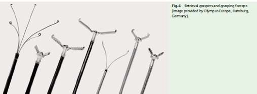
Picture 4. Devices endoscopy depicted from left to right:
Tripronge forceps, alligator forceps, bipronge forceps . 23
Blunt objects usually require immediate endoscopy if they cannot pass through the esophagus perfect, except for coins If the swallowed coin does not show symptoms and is less than 2.5 cm wide, the patient can be observed for the coin. However, if there are symptoms Which critical, like Which happen on patient Which swallow blunt objects, immediate endoscopic removal is required. Objects with a width of more than 2.5 cm or a length of more than 6 cm should also be removed, both for pediatric and adult patients. 14,20
Foreign bodies in the esophagus are an urgent problem, considering that esophagus is structure Which close together with organ vital organs such as the pericardium, aorta, and pulmonary pleura. Disruption of the integrity of the esophageal mucosa can cause serious complications, including leakage of gastric fluid into the mediastinum to perforation. arteries. Attention special required in case bone animals and objects larger than 3 cm, as they can be predictors of perforation. esophagus. Object blunt Which experience clash in esophagus and cause symptom Possible is results from injury ongoing ischemia and requires prompt removal as well. 20 Surgical intervention may be necessary in cases of acute complications such as perforation, occlusion, or hemorrhagic vascular lesions, as well as in cases of failed endoscopic extraction. 13 Prompt surgical consultation and initiation of antibiotics are required if there is perforation with signs of mediastinitis , peritonitis , pneumomediastinum , or pneumoperitoneum . 20
Complications
Complications Which related with object foreign varies depends on the type of foreign object and its condition. In patients who accidentally swallow a foreign object, level complications reported between 3.6% And 7%, without
There have been reports of related deaths. Complications can range from mild mucosal damage to perforation requiring surgery. 12 Mucosal perforation is more likely to occur in patients who has experience >48 O'clock swallowed object foreign or Which have shorter foreign bodies (<7 cm) outside the pylorus. Foreign bodies have a more variable complication rate. The majority of complications This involving injury mucosa light, although There is reports of perforation are approximately 3% in the population with eosinophilic esophagitis. Other types of foreign body ingestion, such as animal bones, dentures, and toothpicks, can have complication rates ranging from 2.8% to 50%. 16
Cases involving the ingestion of bones, especially fish bones, are associated with a higher rate of complications due to predicted injury. mucosa. Factors other Which can predict Complications include delay in intervention of more than 12 hours, sharpness of the object, objects measuring more than 3 cm, location or objects not visible on X-ray. 15
Management of complications depends on their severity. Mild complications that do not involve the gastrointestinal mucosa can usually be managed conservatively by using a nasogastric tube and allowing the area time to heal. Perforation esophagus need Handling Which more directed depending on size disabled, development clinical patient, And presence of comorbidities. Small mucosal perforations or tears can be managed with antibiotics and consideration of covered stent placement. Patients who show signs of sepsis or do not respond to conservative management may require surgical intervention. 20
Esophageal trauma is a condition where damage occurs to the esophagus, like laceration, ulcer or ulceration, perforation, and also rupture or torn wall esophagus. Generally action surgery No treatment is needed for cases of laceration and ulceration of the esophagus. However, if perforation or rupture of the esophagus occurs, this is a very serious emergency situation and can be fatal if not treated properly. Therefore, esophageal perforation is considered the most dangerous event compared to perforation in other parts of the digestive tract. 2,7 Complications that often occur due to foreign objects swallowed in the esophagus are perforation, mediastinitis, fistula or aspirations. 9 Consultation surgery quick required if any of these signs are seen. 20
REFERENCE
Chaudhry SR, Bordeaux B. Anatomy, Thorax, Esophagus . StatPearls Publishing; 2023. https://www.ncbi.nlm.nih.gov/books/NBK482513/?report=printable .
Andrew Su, Parker CH et al. Esophageal anatomy and physiology . Publishing 2020. https://doi.org/10.1016/B978-0-12-813037-7.00005-4 .
Afiaty AS. Esophagoscopy. In: Textbook of Ear, Nose, Throat, Head & Neck Health Sciences. FK UI. Sixth edition. Jakarta; 2007. p. 311 - 313.
Lee KJ, Anatomy of the esophagus. In : Lee KJ, editor, Essential Otolaryngology Head and Neck Surgery. 8th ed. United States of America : The McGraw-Hill Company ; 2013. p. 446- 8.
Schaefer TJ, Trocinski D. Esophageal Foreign Body . StatPearls Publishing; 2023. doi:10.47391/JPMA.04-552.
William W, Shockley. Esophageal Disorders. In : Eibling D, editor. Head and Neck Surgery- Otolaryngology. 4th ed . Philadelphia : Lippincot Williams & Wilkins; 2016: p. 768-70.
Bajwa SA, Toro F, Head A. Physiology, Esophagus . StatPearls Publishing; 2023. https://www.ncbi.nlm.nih.gov/bo oks/NBK519011/?report=printable .
Sahoo L, Padhy RK, Mohapatra D, Ashe S. Foreign Bodies in Upper Gastrointestinal Tract: A Retrospective Analysis of Clinical Intervention and Management. Int J Med Res Prof. 2016;2(5):3–5.
Magalhães-Costa P, Carvalho L, Rodrigues JP, Túlio MA, Marques S, Carmo J, et al. Endoscopic Management of Foreign Bodies in the Upper Gastrointestinal Tract: An Evidence-Based Review Article. GE Port J Gastroenterol [Internet]. 2016;23(3):142–52. Available from: http://dx.doi.org/10.1016/j.jpge.2015.09.002 .
Wang X, Su S, Chen Y, et al. The removal of foreign body ingestion in the upper gastrointestinal tract tract: a retrospective study of 1,182 adult cases. Ann Translation Med . 2021;9(6):1-
8. doi:10.21037/atm-21- 829.
Costa PM, Carvalho L, Rodrigues JP, Tulio MA, Marques S, Carmo J, et al. Endoscopic management of foreign bodies in the upper gastrointestinal tract: an evidence-based review article. GE Port J Gastroenterol. 2016;23(3):142-152.
barrel LI, Ming OF, Ramasamy V, Mohan Singh US, Mohammed I. Transluminal Migration of Oesophageal Foreign Bodies: A Series of Three Patients. J Clin Heal Sci. 2023;8(1):87–92.
Benjelloun N, Idrissi MS, Salihoun M, Acharki M, Serraj I, Kabba N. Foreign Bodies in the Upper Gastrointestinal Tract in Adults: A Reviews of 102 Cases. SAS J Med. 2022;8(4):285– 8.
Long B, Koyfman A, Gottlieb M. Esophageal Foreign Bodies and Obstruction in the Emergency Department Setting: An Evidence-Based Review. J Emerg Med [Internet]. 2019;56(5):499–511. Available from: https://doi.org/10.1016/j.jemermed.2019.01.025
Sobeih A. Extraluminal Perforation Complicating Foreign Bodies in the Upper Aerodigestive. Ann Otol Laryngol. 2010;119:284-8.
Pai K, Pillai S, Bhandarkar A, Anand A, Sabhahit H. Migrating Ingested Foreign Body Of The Upper Aerodigestive Tract with Resultant Septic Shock. Sultan Qaboos Univ Med J. 2013;13:606-10.
Mukherjee D, Banerjee S, Bandyopadhyay S, Mukherjee D, SN B. Extraluminal Migration of Accidentally Ingested Foreign Bodies: an Unusual Case Report. J Evol Med Dent Sci. 2014;3(67):14542–5
Seng LK, Siang LK, Smith JD, Hian YK, Dong F. Complications of Foreign Bodies in the Esophagus. Otolaryngol Neck Surg. 2008;123:613-16.
Lam CK, Woo KS, Van CA. Esophageal Perforation and Neck Abscess from Ingested Foreign Bodies: Treatment and Outcomes. Ear, Nose Throat J. 2008;82:786-94.
John Reyes Genre and Uzma D. Siddiqui. Foreign Body Removal. 2017. 31–41 p.
Birk M, Bauerfeind P, Deprez PH, Häfner M, Hartmann D, Hassan C, et al. Removal of foreign bodies in the upper gastrointestinal tract in adults: European Society of Gastrointestinal Endoscopy (ESGE) Clinical Guideline. Endoscopy. 2016;48(5):489–96.
Maweikere ACF, Mengko SK, Pelealu OCP. Management of Esophageal Foreign Body. e- CliniC , Volume 11, Number 3, September-December 2023, pp. 339-346.
Birch M, The Bauerfeind P, Depressed PH. Removal of Foreign Bodies in The Upper Gastrointestinal Track in Adults: European Society of Gastrointestinal Endoscopy (ESGE)Clinical Guideline. New York. 2016. University of Ulm Albert-Einstein-Allee 23.
afwaf
Comments