FRACTURE MANDIBLE
I Made Nudi Arthana
Fracture mandible is a fracture that occurs in the mandible, which is a bone that forms the lower third of the face and plays an important role in maintaining the shape and contour of the face. The mandible is U-shaped and consists of one corpus. And two fruit hair mandibulae. Mandible support tongue as well as muscle the floor of the mouth and plays a role in the function of mastication, articulation and speech of a person. 1,2 Tissue damage to the mandible can cause various problems ranging from impaired airway patency, problems with mastication function or malcollusion, disorders on articulation talk And swallow, until problem cosmetics. The damage that happen on mandible No only limited on network bone, but may involve defects in soft tissues and adjacent structures. 3 This condition can influence health physique, mentally, And quality life somebody. Mandibular fractures are the second most common facial fractures, because they are the most common bone. Which stand out Which located in edge And his position in one third lower face so it is often the target of forced injuries. In addition, it is a place of attachment of the masticatory muscles so that it has active movement. The diagnosis of mandibular fracture can be indicated by the presence of pain, swelling, tenderness, malocclusion, broken teeth, gaps, uneven teeth, asymmetry of the dental arch, intra-oral lacerations, loose teeth and crepitation. 1,2,3
LITERATURE REVIEW
Anatomy of the Mandible
The mandible is the largest and strongest bone in the face and has a curved shape. resemble letter-U. Mandible is bone flat And consists of from a curved horizontal mandibular body and two mandibular ramus posteriorly. 4,5 The mandibular body and ramus meet to form the mandibular angle (Figure 1). 5
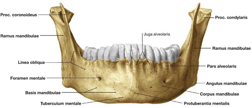
Picture 1. Mandible seen from anterior. 5
The mandibular corpus is divided into the base and the pars alveolaris which are separated by the oblique line which descends from the coronoid process towards the anterior (Figure 2). The pars alveolaris is where the 16 mandibular teeth stand. covers 1 install tooth series central, 1 install tooth series lateral, 1 pair of canine teeth, 2 pairs of premolar teeth, and 3 pairs of molar teeth. 5.6 The mandibular symphysis is located on part base mandible And is line middle meeting between
two parts of the mandible that develop during childhood. Inferior to the symphysis mandible, there is protrusion bone shaped triangle Which shaping the chin Which called protuberantia mentalist. On part inferior tooth premolar both are there foramen mental Which is place the walk nerve mentalist and blood vessels. 5,6
Each – each hair mandible own One Process coronoid and the condylar process. Through the condylar process, the mandible articulates with the bone temporal on joints temporomandibular (TMJ) Which allows the opening and closing movements of the mouth for mastication. The movement of the mandible is facilitated by four masticatory muscles including the masseter, temporalis muscle, pterygoid medialis And muscular pterygoid lateral. Fourth These muscles are innervated by the mandibular division of the trigeminal nerve (V3). These muscles have insertions on the ramus of the mandible and the coronoid process and the condylar. Muscle pterygoid lateral facilitate motion opening the mouth and protrusion of the mandible, while the other three muscles facilitate the closing and elevation movements of the mandible. Disruption of the attachment of the masticatory muscles can disrupt the balance and coordination of muscle movements, causing pain and disruption of the mouth opening movement. 4,5,6
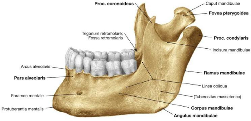
Picture 2. Mandible from side lateral left. 5
The temporomandibular joint is a bilateral diarthrodial joint (there are two component articulation bone) And joints synovial And own discus articular in its capsule. The movement of the TMJ resembles a hinge and is useful in mastication and articulation during speech. The TMJ capsule is a fibroelastic structure and is innervated by the auriculotemporal nerve . The TMJ articular disc separates the condyle and articular fossa of the temporal bone. The smooth surface of the disc allows free movement of both bony surfaces. 4,6
On inner side of the ramus mandibulae, there appears to be a tiered crest or Linea mylohyoidea which is the attachment point for the mylohyoideus muscle which limits the height of the floor of the mouth. The mental spine is located on the posterior aspect of the symphysis, above the linea mylohyoidea. The upper part of the mental spine is the attachment point for the genioglossus, while the lower part is the attachment point for the adhesion geniohyoid. Spina mentalist can No found or merge and form an elevation in some individuals. The lingular foramen (above and/or below the mental spine) and the lateral lingular foramen (in the premolar area) can be found in most mandibles. Both foramina are the entry points for blood vessels for the symphysis. On the inner aspect of the mandibular angle, the pterygoid tuberosity is found. 4,6
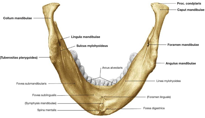
Picture 3. Mandible from looks inferior. 5
On the posterior aspect of the ramus, the mandibular foramen can be found (Figure 3). This foramen is where the alveolar neurovascular canal begins. inferior. Lingula mandibulae is A protrusion bone Which becomes the attachment site for the sphenomandibular ligament. The mandibular lingula is a marker for local anesthetic injection in the form of an inferior alveolar block. 5,6
The date tooth, specifically on carry on age can cause regression pars alveolaris mandibulae. Regression can to be continued until forame mental located at the top of the mandible In some cases (Figure 4), the mental foramen may disappear, causing the maxillary nerve to be injured. The angle of the mandible has a larger angle than the mandible and teeth. 5
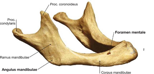
Picture 4. Mandible elderly . 5
The blood supply to the corpus mandibula comes mainly from the inferior alveolar artery. as well as from vessels blood Which supply m. genioglossus, m. geniohyoid and anterior belly of digastric muscles (Figure 5). These three blood vessels receive blood supply from the sublingual branch of the lingual artery and the facial branch of the submental. Branch – branch from vessels blood the can perforates the lingual cortical plate and anastomoses with vessels from the inferior alveolar artery. Branches of the submental artery can anastomose with the mental artery and provide a retrograde blood supply to the body and symphysis of the mandible. The ramus of the mandible and the angle of the mandible are supplied by the inferior alveolar artery which also supplies blood to m. masseter And m. pterygoid medial. Flow blood for the coronoid process comes from the blood vessels that supply the temporalis. 5,6 The innervation of the mandible comes primarily from the mandibular nerve which is branch from nerve cranialis (CN) trigeminal (V3). Nerve The mandibular nerve has anterior and posterior divisions. In the anterior division there is the mandibular nerve. buccalis and nerve branches motor innervates the lateral pterygoid, m. masseter, and m. temporalis. While in the posterior division there are branches of the lingual nerve, the inferior alveolar nerve, and the auriculotemporal nerve. The mental nerve is a branch of the inferior alveolar nerve. 4,5,6 The inferior alveolar nerve is located behind and more lateral to Lingual nerve. This nerve supplies sensation to the teeth and mandible. The inferior alveolar nerve exits the mental foramen as the mental nerve fibers, and supplies sensation to the labial gingiva, mucosa, and lower lip/chin. The lingual nerve exits beneath the lateral pterygoid and is closely related to the lingual aspect of the mandible. It contains taste fibers for the anterior 2/3 of the tongue and preganglionic parasympathetic fibers for the submandibular ganglion (Figure 6). 5
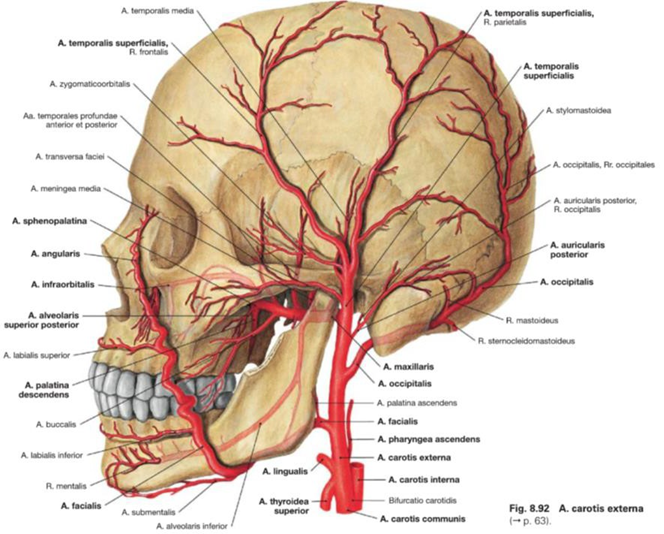
Picture 5. Vessels blood mandible. 5
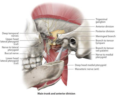
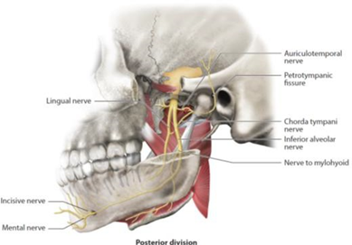
Picture 6. Mandibular nerve anterior division (left) And posterior (right). 5
Definition And Classification Mandibular Fracture
Fracture is defined as a linear deformity or discontinuity of bone caused by force. Fractures can occur due to trauma or due to pathological processes. Mandibular fracture is the breakup continuity of the mandibular bone. 3.7 Mandible is bone Which strong, but on a number of where the weak part is found. Corpus area mandible especially consists of dense cortical bone with little substance spongy as where blood vessels pass And vessels lymph. Thin area on The mandible is the angle and subcondyle so that this part is included in the weak part of the mandible. In addition, weak points are also found in the mental foramen, the mandibular angle where the molar III teeth erupt, especially the condylar column mandible especially when trauma from front direct about chin then the force will be transmitted backwards. 3.8
Fracture lines in the mandible usually occur in weak areas. of the mandible depending on the trauma mechanism that occurs. The subcondylar fracture line is generally below the neck of the condyloid process due to fighting and is almost vertical. However on accident Then cross-line fracture happen near with condylar caput, the fracture line that occurs is oblique. 7 In the angle region The fracture line is generally below or behind the third molar region towards the mandibular angle. On fracture corpus mandible line fracture No always parallel with axis tooth, often line fracture shaped oblique. Line fracture started on
The alveolar region of the canine and incisors runs obliquely towards the midline. 7,8 In mandibular fractures, the fractured fragment is displaced. due to the pull of the masticatory muscles, therefore reduction and fixation of mandibular fractures must use splinting to counteract the pull of the masticatory muscles. Several factors that influence mandibular fracture displacement include: direction and strength trauma, direction And corner line fracture, There is or no teeth on fragments, direction of muscle detachment and extent of damage soft tissue. In the mandibular ramus area seldom happen fracture, Because area This fixed by muscular master on part lateral And medial bymusculus pterygoid medial. Thus also on the coronoid process which is fixed by the masseter muscle. 3,7,8
A number of types of classification fracture mandible can classified based on:
Incident fracture mandible in accordance with location its anatomy; process condyloideus (29.1%), angle mandible (24%), symphysis mandible (22%), corpus mandible (16%), alveoli (3.1%), hair (1.7%), process coronoid (1.3%). 3
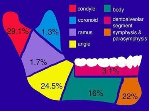
Picture 7. Region mandible And frequency fracture mandible based on region 3
2. Based on There is whether or not tooth on left And right line fracture; class I: teeth are present on both parts of the fracture line, class II: teeth are present on only one part of the fracture line, class III: no teeth on both fragments, perhaps the teeth were previously absent (edentulous) or teeth were lost during the trauma. 7,8
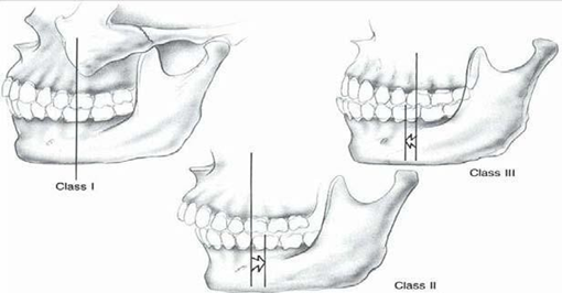
Picture 8. Relationship There is whether or not tooth on line fracture. 3
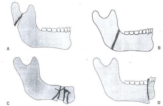
3. Based on the direction of the fracture and ease of repositioning, it is distinguished: horizontal And vertical Which shared become favorable And unfavourable . The criteria for favourable and unfavourable are based on the direction of a fracture line to the muscular force acting on the fragment. It is called favourable if the direction of the fragment makes it easy to reduce the bone during reposition, while unfavourable if the fracture line makes it difficult to reposition. 3,7
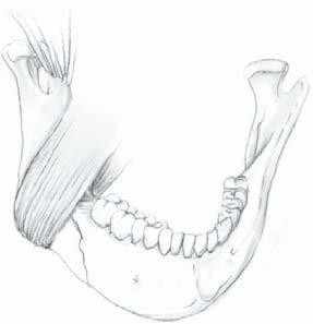
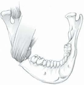
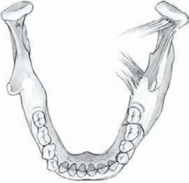
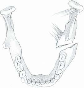
Figure 9. A. Horizontal favorable fracture , B. Horizontal unfavorable fracture, C. Vertical favourable fracture , D. Vertical unfavourable
4. Based on the weight degrees fracture, shared become fracture simple or closed , that is, without any connection to the outside world and no discontinuity of the tissue around the fracture. Compound or open fractures, that is, fractures relate with world outside Which involving skin, mucosa or periodontal membrane.
5. Based on the type of fracture, it is divided into greenstick or incomplete fractures; an incomplete fracture where one side of the bone is fractured while the other side of the bone is still attached. Greenstick fractures are usually found on children Because periosteum thick. Fracture single; fracture only in one place. Multiple fractures; fractures Which occurs in two or more places, generally bilateral. Comminuted fracture; there are small fragments that can in the form of simple fracture or compound . Besides That there is Also fracture pathological; fractures that occur due to metastases to the bone, impacted fractures; fractures with one of the fracture fragments inside another fracture fragment. Atrophic fractures ; are spontaneous fractures that occur in atrophic bones such as in the jaw that has no teeth. Indirect fractures; fractures that occur far from the site of trauma. 7,9
Picture 10. Type fracture mandible. A. Greenstick ; B. Simple ; C. Commuter ; D.
Compound 3
Epidemiology Fracture Mandible
Fracture on midface often happen consequence accident vehicle motorized, fall, violence And consequence trauma object blunt other. Based on research by Natu et al , mandibular fractures occur more frequently in men (81.8%) than women (18.2%) with percentage highest on age 21-30 years (28.8%), followed by age 11-20 year (25.8%). Accident Then cross is the most common cause of fractures, followed by falls from a height. As many as 56.1% fracture is fracture unilateral And 62.1% is fracture isolated in the mandible where the parasymphysis (31.4%) is the most frequent fracture location in the mandible followed by the body (24.5%). 7
Biomechanics Mandible
Mandible own mobility And style Which very Lots, so that In handling mandibular fractures, the biomechanics that occur must be really considered. Mandibular movement is influenced by four pairs of muscles called the chewing muscles, namely: the masseter, temporalis, lateral pterygoid and medial muscles. 3 When opening the mouth, the inferior lateral pterygoid muscle contracts, followed by the superior lateral pterygoid muscle when opening the mouth wider. Meanwhile, the role of closing the mouth is the temporalis and masseter muscles, reinforced again by the medial pterygoid muscle. 3,9
Etiology Fracture Mandible
A hard blow to the face can cause a mandibular fracture. Tolerance mandible to clash more tall than other facial bones. Mandibular fractures are more common than facial bone fractures. Which other Because form mandible Which stand out so that sensitive against impact. In general, mandibular fractures are caused by direct trauma. 1,3,10
Mandibular fractures can be caused by trauma or pathological processes. According to Kruger, 69% from fracture mandible due to by violence physical, 27% accidents, 2% due to sports and 4% pathological factors, while pathological fractures can be caused by cysts, bone tumors, osteogenesis imperfecta, osteomyelitis, osteoporosis, atrophy or bone necrosis. 7,11
Diagnosis Fracture Mandible
In establishing the diagnosis of mandibular fractures, including anamnesis, if it is a case of trauma, the trauma mechanism or mode of injury must be known , physical examination and supporting examination. 3,4,7 In cases of trauma, examination of patients with suspected mandibular fractures must follow the ATLS ( Advanced Trauma Live Suport) rules, which consist of an initial examination or primary survey which includes examination of the airway, breathing, circulation and disability . In trauma patients with mandibular fracture must be noticed existence possible airway obstruction which can be caused by the mandibular fracture itself or by intraoral bleeding which causes blood aspiration. 4
After the primary survey has been conducted and the patient's condition is stable, a secondary survey examination can be continued , including: Anamnesis, in the anamnesis subjective complaints related to mandibular fractures are suspected from the presence of pain, swelling, abnormal occlusion, numbness in the distribution of the mental nerve, swelling, bruises, bleeding from socket tooth, tooth Which fracture or date, trismus, inability chew. Besides That complaint usually accompanied by history trauma like accident Then traffic, violence, falls, sports accidents or a history of pathological diseases.
In general, maxillofacial trauma can be identified during an initial examination or primary survey or secondary examination or secondary survey . 4 Examination of the respiratory tract is important because trauma can just cause disturbance road breath. Blockage can caused by the tongue the fall tongue toward behind, can also by closed channel breath due to mucus, blood, vomit and foreign objects. Local examination of mandibular fractures, between; extraoral clinical examination, above the fracture site there is usually ecchymosis and swelling. Soft tissue lacerations often occur and deformation of the bony mandibular contour can be clearly seen. If there is displacement of the fragments the patient cannot close teeth front And mouth hang slack And open. Patient often seen support jaw lower with hand. Can also water saliva mixed blood dripping from corner mouth patient. Palpation gentle with ends finger performed on the condyle area on both sides, then continued along the lower border of the mandible. Softened parts should be found in fracture areas, as should changes in contour and bone crepitation. 4.7
Inspection clinical intraoral, every tooth chips Which broken must removed from the mouth. The buccal sulcus is examined for ecchymosis and then the lingual sulcus. Hematoma in the lingual sulcus due to mandibular trauma almost always pathognomonic of mandibular fractures. 4.7 Supporting examination; in mandibular fractures can done inspection support between other; Photo X-ray to determine the fracture pattern that occurs. Every radiological examination is expected to produce image quality that includes the area being observed, namely the pathological area and the surrounding normal area. Eisler Photo, This photo was taken for imaging of the mandible. ramus and corpus, made on the right side or left as needed. Town′s view ; made to see the projection of the maxillary bone, zygoma and mandible. Reverse Photo Town's view ; done For see existence mandibular condylar neck fractures especially those displaced medially and can also be used to see the lateral wall of the maxilla. Panoramic photo ; also called pantomography or rotational radiography madefor know condition mandible start from the right condyle to the left condyle along with the position of the teeth including occlusion of the tooth maxilla. Profit panoramic is; coverage anatomical Which area, radiation dose that low, examination is quite comfortable, can be done on trismus patients. The disadvantage is that it cannot show a clear anatomical picture of the periapical area as produced by intraoral photos. Temporomandibular Joint; in patients with direct trauma, the chin area is often found condition on chin Good, will but happen fracture on area mandibular condyle so that the patient complains of pain in the TMJ area when opening the mouth, trismus sometimes a little malocclusion. In making standard TMJ photos, it is usually done projection lateral open mouth or Parma and projection lateral normal or Schuller mouth closure. Orbitocondylar view ; performed to view the TMJ when opening the mouth. mouth wide, show condition structure And contour from bald condyle seen from the front. 8. CT Scan; This examination is performed in emergency cases. Not yet is inspection standard. CT Scan especially For fracture maxillofacial Which very complex. 8,10
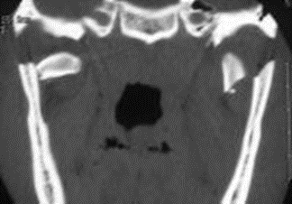
Picture 11. CT Scan coronal fracture bilateral condyles . 8
Management Fracture Mandible
Principle Handling fracture mandible on step beginning emergency such as road breath or airway , breathing or breathing , blood circulation including treatment shock or circulation , Handling wound network soft And temporary immobilization and evaluation of possible brain injury. The second stage is definitive fracture management. Mandibular fracture management is generally divided into two methods, namely closed and open reposition. In closed reposition or conservative , reduction fracture And immobilization mandible achieved by placing maxillomandibular fixation equipment. Open reposition of the fractured part is opened surgically, the segment is reduced and fixed directly using wires or plates called wire or plate osteosynthesis . Technique open And closed No always done alone, but sometimes- sometimes combined. Approach third is is modification from technique open that is method fixation skeletal external. On management mandibular fracture always be noticed principles dental And orthopedicsso The fractured area will return to or approach its true anatomical position and good masticatory function. 3,4,7
Closed reduction of lower jaw fractures, namely conservative treatment by performing repositioning without direct surgery on the fracture line and performing immobilization with interdental wiring or external pins. fixation . Indication For closed reduction between other: a. fracture communicative as long as the periosteum is still intact, bone healing can be expected, b. fractures with damage soft tissue Which Enough heavy Where reconstruction soft tissue can be used rotation flap And free flap when wound the No too big. c. edentulous mandible, d. fracture in children, e. fracture condyle. Technique which is used in mandibular fracture therapy closed reduction is intermaxillary fixation. Fixation This maintained 3-4 Sunday on fracture area condyle and 4-6 Sunday on area other from mandible. Profit from reposition closed is more efficient, has lower complication rates and shorter operating times. This technique can be performed at the outpatient level. Disadvantages include fixation Which long, disturbance nutrition, risk ankylosis TMJ or temporomandibular joint and airway problems. 4,9,12 Some intermaxillary fixation techniques include:
Eyelet or ivy loop technique , placing an ivy loop using a 24-gauge wire between two stable teeth using a smaller wire to give fixation maxillomandibular (MMF) between loops ivy. The advantage of this technique is that the material is easy to obtain and causes minimal damage to periodontal tissue and the jaw can be opened by simply lifting it. bond intermaxillary. The disadvantages wire easy separated time used for intermaxillary fixation, 9,11
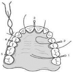
Picture 12. Technique eyelet or ivy loop.9
Arch bar technique , Indications for installing an arch bar are missing or missing teeth. sufficient for installation of other methods, accompanied by maxillary fractures and fragments obtained dentoalveolar on Wrong One end jaw Which need to be reduced according to the jaw arch before intermaxillary fixation is installed. The advantages of using an arch bar are easy to obtain, low cost, easy adaptation and application. The disadvantages are that it causes inflammation of the gingiva and periodontal tissue, and cannot be used in patients with extensive edentulousness. 9,11
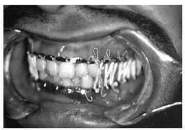
Picture 13 . Fixation maxillomandibular. 9
Reposition open (open reduction) ; action operation For correcting malocclusion deformities that occur in lower jaw fractures by performing direct fixation using wire ( wire osteosynthesis ) or plate ( plate osteosynthesis ) . Indication For reposition open reduction : a. displaced unfavourable fracture through the angle, b. displaced unfavourable fracture of the corpus or parasymphysis, c. multiple facial bone fractures, d. midface fracture accompanied by bilateral displaced condylar fractures. The open reduction surgical technique is a type of clean contamination surgery, requiring general anesthesia. The advantages of open reduction include: earlier mobilization and better reapproximation of bone fragments. The disadvantages are that it is more expensive and requires an operating room and anesthesia for the procedure. 4,8,9
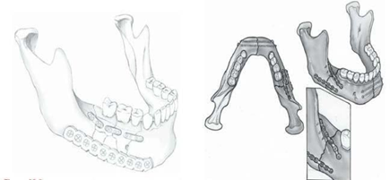
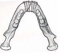
Picture 14. Technique operation reposition open (open reduction). 3
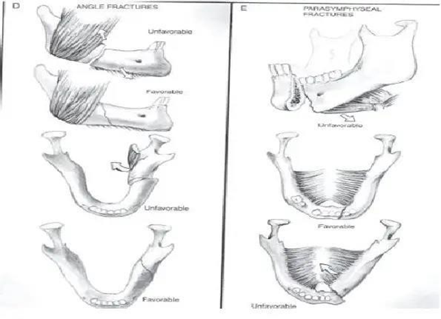
Picture 15 . Favorable fracture and unfavorable. 12
Open reduction of mandibular fractures has intra and extraoral approaches. Extraoral approaches can be performed via the submandibular, submental, or preauricular.
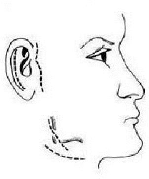
Picture 16. Extraoral approach
Picture 17. Incision Mandible. 12
With the intraoral approach, the mandibular region is reached through a vestibular incision in the mucosa. Compared with the extraoral approach, the intraoral approach is faster, has no extraoral scars, and has a lower risk of affecting the facial nerve. The materials that can be used in open reposition include wire, wire mesh , plates and screws , and others. 12
Wiring (wire)
The wire is made like an eye, then an eye earlier installed around two teeth or molars in the upper or lower jaw. The broken lower jaw is fixed to the upper jaw through the eyes on the upper and lower wires. If necessary, these wire ties are installed in various places to obtain strong fixation.
Plating
Plate installation aims to give prisoner on fracture area, so that can unite part fracture with alveoli superior. After the plate is installed, maxillary fixation is no longer needed. Note that the screw installation on the plate is not done with too much pressure. Because with installation screw Which too strong will resulting in the occurrence of difficulty on moment release,by Because That, installation with a technique that does not apply too much pressure is preferred in installing plates on mandibular fractures. Follow-up after surgery is to provide analgesic as well as give antibiotics spectrum wide on fracture patients and evaluated for nutritional needs, monitor intermaxilla fixation during 4-6 Sunday. Tighten up cable every 2 Sunday. After wire is opened, evaluate with panoramic photo to ensure the fracture has united. 9,12
Complications
Complications after repair of mandibular fractures are generally rare. The most common complication of mandibular fractures is infection or osteomyelitis which can then can cause various other complications. The mandible is the area that most often experiences fracture healing disorders, both malunion and nonunion. Complaints given can be in the form of prolonged pain and discomfort in the jaw joint or temporo mandibular joint due to changes position And instability inter-joint jaw left And right.Thing This not only affects the joints but the chewing muscles and muscles around the face can also provide a pain response. 9,10
There are several risk factors that are specifically related to mandibular fractures and have the potential to cause malunion or nonunion. The biggest risk factors are infection, then poor apposition. Good, lack of immobilization segment fracture, existence foreign object, pull unfavorable muscle tone in the fracture segment. Severe malunion of the mandible will resulting in facial asymmetry and can also be accompanied by functional disorders. These abnormalities can be corrected by planning osteotomies appropriately to reconstruct the shape of the mandibular arch. 9
Maintenance after fixation or immobilization
After do maintenance mandibular fracture with reposition, fixation and immobilization to be continued with maintenance; maintenance health general includes giving antibiotics, analgesics, roborantia and nutritious food, maintaining oral hygiene , maintenance fixation tools, and perform physiotherapy. Follow up after done operation is with give analgesic as well as give broad spectrum antibiotics to fracture patients open and evaluated nutritional needs, monitor intermaxilla fixation for 4 - 6 Sunday. Tighten up wire every 2 weeks. After wire opened, evaluation with Photo panoramic For ensure fracture has union.4,8,9
CONCLUSION
Mandibular fractures are the second most common facial area due to their prominent position and location in the lower third of the face. The main goal of mandibular fracture treatment is to restore chewing and speech function. Matter This can achieved with election modality Which appropriate, technique correct operation especially in achieving mandibular occlusion, as well as post-operative care and rehabilitation. In the management of mandibular fractures It is necessary to understand the biomechanics of the mandible so that the correct fixation position can be estimated and satisfactory results can be obtained.
REFERENCE
Zhang Q, Wu W, Qian C, Xiao W, Zhu H, Guo J, et a. Advanced biomaterials for repairing and reconstruction of mandibular defects. Mater Sci Eng C. 2019;103(February):109858.
Krüger E. Chicago: Quintessence Publishing Company; 2010. Mandibular fractures, 1. Classification, diagnosis and fundamentals of treatment; pp. 211– 223.
Jonas T. Johnson, Clark A. Rosen. Mandibular Fracture in Bailey's Head and Neck Surgery. Fifth Edition. 2014. P. 1229-1241.
Brennan PA, Mahadevan V, Evans BT. Clinical Head and Neck Anatomy for Surgeons. Clinical Head and Neck Anatomy for Surgeons. 2015. 1–324.
Brennan PA, Mahadevan V, Evans BT. Clinical Head and Neck Anatomy for Surgeons. Clinical Head and Neck Anatomy for Surgeons. 2015. 1–324 p.
Paulsen F, Waschke J. Head. In: Sobotta Atlas of Anatomy Head, Neck and Neuroanatomy. 18th ed. Munich: Elsevier; 2018. p. 4–110.
Natu SS, Pradhan H, Gupta H, Alam S, Gupta S, Pradhan R, Mohammad S, Kohli M, Sinha VP, Shankar R, Agarwal A. An epidemiological study on pattern and incidence of mandibular fractures. Plastic surgery international. 2012;2012.
Robert E. Lincoln. Pratical Diagnosis and Management of Mandibular and Dentoalveolar Fracture in Facial Plastic, Reconstructive and Trauma Surgery. 2004. P. 597-627.
Pickrell BB, Cerebral AT, Maricevich Hospital. Mandible fractures. In Seminars in plastic surgery 2017 May (Vol. 31, No. 02, pp. 100-107). Thieme Medical Publishers.
Panesar K, Susarla SM. Mandibular Fractures: Diagnosis and Management. InSeminars in Plastic Surgery 2021 Oct 11 (Vol. 35, No. 04, pp. 238-249). 333
Seventh Avenue, 18th Floor, New York, NY 10001, USA: Thieme Medical Publishers, Inc. Good day RHB. Management of fracture of the mandibular body and symphysis. Oral Maxillofac Surg Clin North Am. 2013 : 25 (4) : 601 – 16.
Sadhwani BS, Anchlia S. Conventional 2.0 mm miniplates versus 3-D plates in mandibular fractures. Annals of maxillofacial surgery. 2013 Jul;3(2):154.
Comments