Juvenile nasopharyngeal angiofibroma
I Made Nudi Arthana
Juvenile nasopharyngeal angiofibroma (JNA) is a benign vascular tumor located on part posterior nasopharynx And foramen sphenopalatine. JNA very much rare where incident tumor This only covering 0.05% until 0.5% from all over tumor head and neck. The prevalence of JNA in the United States is estimated at only 0.4 per million population. Although JNA can happen on all age, However incident JNA highest found in male patients aged 7 to 29 years old. 1.2 Therefore disease This quite rare, no data related to JNA epidemiology was found in Indonesia.
LITERATUR REVIEW
Anatomy Nasopharynx
The nasopharynx is the most superior space of the pharynx. The nasopharyngeal space is cuboidal in shape and is bounded by the cranial base on the superior side, the soft palate on the inferior side, the nasal cavity on the anterior side, the posterior pharyngeal wall on the posterior side, and the pterygoid plates madeial And muscle constrictor pharynx on side medial. Nasopharynx has an anteroposterior diameter of about 2 cm with a height of about 4 cm. In the nasopharynx, several structures can be found such as the eustachian tube opening, torus tubarius, fossa rosenmuller, and adenoids. The eustachian tube opening is located on the posterolateral wall of the nasopharynx. The torus tubarius is located posterior to the eustachian tube opening, while the fossa rosenmuller is located supero-posterior to the torus tubarius, adenoids are located on the upper and posterior walls of the nasopharynx (Figure 1). 7
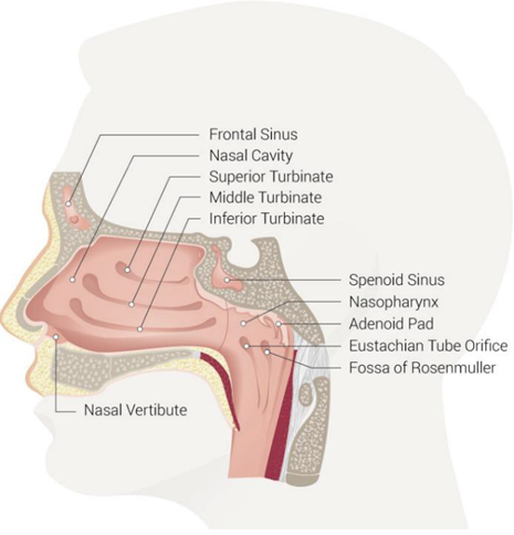
Picture 1. Anatomy Nasopharynx. 7
The nasopharynx has various functions, including respiratory function, immunity, equilibrium, until production voice. Nasopharynx functioning as Wrong One channel upper respiratory tract that connects the nasal cavity with the larynx and trachea through the oropharynx. The nasopharynx also plays a role in the immunity process where debris from the filtered air will be transported out from body through mucociliary mechanism clearance to the oropharynx. Besides That, on nasopharynx Also there is adenoids Which play a role catch bacteria and dirt that enter through the oral cavity and prevent them from entering the respiratory tract. The muscles in the nasopharynx (tensor veli palatini, levator veli palatinin, and salphingopharyngeus muscles) also play a role in the process of equilibrium or body balance by controlling the opening and closing of the eustachian tube opening so that the pressure between the middle ear and nasopharynx is equal. The tensor veli palatini and levator veli palatini muscles also function to cause contraction and elevation of the soft palate which prevents food from the oropharynx from entering the nasopharynx during the swallowing process. 7
The main blood supply of the nasopharynx is provided by the mandibular artery which is branch from artery carotid internal, artery maxillary (artery pterygoplatina), facial artery ( ascending palatine artery ), and ascending pharyngeal artery (superior pharyngeal artery). Venous drainage from the nasopharynx is via the parapharyngeal venous plexus. This venous plexus then drains further into the retropharyngeal and facial veins which then into the internal jugular vein. Lymphatic drainage from the nasopharynx first leads to lymph gland in the retropharyngeal area where these glands are known as Rouvière . Furthermore, lymphatic drainage from the retropharynx will lead to the internal jugular lymph nodes. 7
Innervation sensory from nasopharynx front especially done by nerve maxillary which is branch from nerve trigeminal (CN V2), whereas innervation sensory from the posterior nasopharynx is mainly carried out by the glossopharyngeal nerve (CN IX). The motor innervation of the muscles in the nasopharynx comes from the vagus nerve (CN X), except For muscle tensor veli palatine Which innervated by nerve mandibular (CN V3). 7
Definition JNA
Histologically, JNA is defined as an irregular vascular lesion enclosed in a pseudocapsule composed of numerous blood vessels of varying sizes embedded in a fibrous stroma rich in collagen and fibroblasts. This vascular lesion is benign, but invasive. 8,9
Epidemiology JNA
JNA is one of the very rare benign tumors of the head and neck. JNA is estimated to only cover 0.05% to 0.5% of tumor cases in the head and neck area. Prevalence JNA in all over world estimated span from 1 per 150,000 until 1 per 1,500,000 population. This tumor tends to be more common in the male population aged 7 - 29 years. This predominance is thought to be related to hormonal factors, especially androgens, Which reach the peak on population teenager male . 4.10
Etiopathogenesis JNA
The exact etiopathogenesis of JNA is still unknown to date. Until now This there is a number of theory related etiology JNA, covering theory angiogenesis, hormonal, histogenetic, and viral infections.
Anomaly vascular
JNA is thought to originate from vascular anomalies, such as arteriovenous malformations, which originate from the remnants of the first branchial arch. The regression process is incomplete in the area around the foramen. sphenopalatine will cause formation remainder network vascular from the first branchial arch which is thought to be the beginning of the formation of JNA . In addition, it is thought that in patients with JNA there is an increase in the expression of angiogenesis factors and growth factors that cause the development of the remaining first branchial arch arteries into a vascular mass. Several angiogenesis factors and growth factors found to be increased in patients with JNA include vascular endothelial growth factor (VEGF), Flt-1, fibroblast growth factor (FGFR), platelet-derived growth factor (PDGF), as well as Hypoxia Inducible Factor -1α (HIF-1α). Improvement these potent angiogenic factors also mentioned stimulated by androgen hormones so that anomaly theory vascular This allegedly related with theory hormonal from pathogenesis JNA. 3,4,11
Hormonal
Androgens are thought to play a role in the pathogenesis of JNA due to the predominance of young males in JNA cases. In this theory, JNA is considered a fibrovascular tumor that develops under the influence of hormonal fluctuations during puberty. In addition to androgens, lower estrogen expression is also allegedly play a role in pathogenesis JNA. Matter This supported with existence consistent findings in various studies related to increased androgen receptor expression and decreased estrogen and progesterone receptor expression. In addition, several studies showed a 44% decrease in tumor size in JNA patients who received anti-androgen therapy, namely flutamide. 3,11
Genetics
The presence of male predominance and the malignant transformation process of JNA in some patients has led to the suspicion of a genetic role in JNA, but the role of this genetic factor in the pathogenesis of JNA is still unknown. Several previous studies have mentioned a link between genetic mutations and glutathione-S-transferase M1- null (GSTM1- null ) genotype polymorphism , C-myc oncogene, and Kit proto-oncogene receptor tyrosine kinase (c-Kit). GSTM1- null polymorphism can cause somatic mutations of β-catenin or germline APC mutations that act as co-activators of androgen receptors. Thus, the presence of GSTM1- null polymorphism will increase the potential from Work androgen hormone. C-myc oncogene and c-Kit able to increase angiogenesis activity by increasing the release of growth factors in stromal cells, thereby triggering the development of JNA. 3,11
Infection virus
Human Papilloma Virus (HPV) and Epstein-Barr Virus (EBV) infections are thought to be associated with the pathogenesis of JNA because infection with these two viruses has been known to have a tumorigenic effect on various cases of other neoplasms, such as Kaposi's sarcoma. And lymphoma Hodgkin . A number of studies previous mainly reporting the presence of HPV findings in JNA tissue so that it is suspected that HPV infection can accelerate the proliferation of stromal cells in JNA. 3,11
Pathogenesis-wise, there are several theories related to the origin and expansion of JNA. JNA was initially thought to originate from the nasopharyngeal area, so it was called nasopharyngeal angiofibroma. However, along with the development of studies, there are several reports that JNA can originate from the sphenopalatine foramen / pterygopalatine fossa, the canal vidianus, koana, nasopharynx, as well as pterygoid wedge. Expansion lesi JNA Also can occur to various directions so that can cause symptom Compressive Which varies depending on the location of origin and extent of the JNA lesion (Figure 2).
a. Foramen sphenopalatine or fossa pterygopalatine
Most studies state that the sphenopalatine foramen or pterygopalatine fossa is the origin of JNA (48% of JNA cases). JNA originating from this area is more likely to experience medial and lateral expansion because area the own more A little prisoner. Distribution JNA to direction lateral will cause tumor invasion towards the infratemporal fossa through the pterygomaxillary fissure and to direction cheek. Distribution of lesions JNA to direction medial can cause invasion tumor to nasopharynx And cavity rice, including sine maxillary And ethmoid. Tumor on This area can also extend posteriorly, causing invasion of the vidian canal, parapharynx, and pterygoid processes. 3,9
Channel vidianus
The vidian canal is often considered as one of the sites of origin of JNA based on radiological findings and the theory that JNA lesions originate from the remnants of the first branchial arch. However, a systematic review study reported that only 0.4% of JNA cases were suspected to involve the vidian canal. The vidian canal is a bony duct located between the pterygoid process and the sphenoid sinus that connects fossa pterygopalatine with foramen lacerum. Lesion JNA Which originating from the vidian canal is thought to be able to invade surrounding structures such as the choana, nasal cavity, pterygopalatine foramen, pterygopalatine fossa, and nasopharynx. 3,9
Koana and nasopharynx
Another theory states that JNA lesions originate from the choana and nasopharynx where the tumor will then spread towards the pterygoid and sphenoid bones as the tumor increases in size. 3,9
Pterygoid wedge
Wrong One studies mention that pterygoid wedge is Wrong One the most consistent location of JNA origin due to the finding of pterygoid wedge involvement in 99.1% of 242 JNA cases based on pre-operative radiological examination. 3,9

Picture 2. Scheme Direction Expansion Typical from JNA. a) expansion median from JNA originating from the sphenopalatine foramen and b) the paramedian extension of the JNA originating from the pterygopalatine fossa. 12
Diagnosis JNA
The diagnosis of JNA is generally made based on the presence of symptoms due to obstruction from mass JNA Which invade or obstruct structure anatomical around Which followed by the finding of a vascular mass in the nasopharynx, choana, vidian canal, or other structures based on physical examination and radiological examination.
Anamnesis And Inspection Physique
A common symptom found in JNA is the presence of progressive unilateral nasal obstruction. Which marked with existence nose dead end, (100%), rhinorrhea (80-90%) and epistaxis unilateral recurrent (75%). Symptoms and signs others that can found at JNA is painful head And painful on face (75%) Which happen consequence obstruction paranasal sinuses or otitis media secretory repetitive Which happen Because disturbance function Eustachian tube due to obstruction by JNA mass. Suspicion of JNA becomes stronger if these symptoms are found in young male patients. As the mass develops, JNA can cause significant facial swelling (75%), hearing loss and ear pain (37.5%), proptosis (37.5%), changes in tongue position (25%), and even visual impairment (12.5%). 6,8,9 paresis nerve cranial II, III, IV And VI generally will found if the mass has invaded the infratemporal fossa.
Inspection Endoscopy Nasal Nasal endoscopic examination generally shows a lobulated mass . is at in behind concha media so that ta m b pack fulfil koana. Mass those found generally have a smooth surface and appear hypervascular (Figure 3).

Picture 3. Inspection Endoscopy Nasal on Case JNA. a) Endoscopy on The right nasal cavity shows a smooth-surfaced mass originating from the back of the middle concha with total obstruction of the nasal cavity; b) Nasal endoscopy on the contralateral side shows an JNA mass filling the rhinopharynx and causing deformity in the posterior part of the nasal septum. 8
Inspection Radiological
Radiological examination plays an important role in establishing the diagnosis and determining the stage of JNA. Radiological examinations that are generally recommended for JNA cases include Computed Tomography (CT) -Scan examinations. head or Magnetic Resonance Imaging (MRI) head. Inspection X-ray is no longer used to diagnose JNA. CT scans generally show a lobulated , heterodense, non-capsulated soft tissue mass of varying size (depending on tumor progression) that is usually found centered in the pterygopalatine fossa. JNA generally involves nasopharynx And fossa pterygopalatine so that cause widening from the sphenopalatine foramen and anterior bowing of the posterior wall of the maxillary sinus ( Holman-Miller sign ). CT scan of the head in advanced JNA will show involvement of the sphenoid, maxillary, or etomidal sinuses, as well as the nasal fossa, infratemporal fossa, orbit, or skull base. CT scan of the head with contrast will show enhancement indicating increased vascularity in the JNA mass (Figure 4). 8,9,13

Picture 4. Inspection CT Scan Head on 2 patient JNA different. a) JNA unilaterally on the left side is small in size originating from the foramen
sphenopalatine. b) JNA recurrence on nasopharynx. 8
MRI examination of the head may not be as good as CT- Scan examination in showing changes or bone erosion caused by JNA invasion, but MRI examination of the head is very important in cases of advanced JNA. suspected to have caused intracranial invasion (through the skull base or orbital wall). MRI examination of the head generally shows a heterogeneous mass with various signal flows ( flow-voids ) due to the many blood vessels in the mass which are characterized by a salt and pepper image. MRI T1- weighted in cases JNA show intensity similar with intensity muscle, whereas on MRI T2- weighted generally looks existence hyperintense. Use contrast on MRI will show the presence of stinging in either the T1 or T-2 sequence (Figure 5). 8,9,14

Picture 5. Inspection MRI head with contrast piece (a) axial And (b) coronal showing the presence of an JNA mass which is thought to originate from
process pterygoid left Where lesi This has invade nasopharynx (asterisk) black) and compartments intracranial extradural through fissure inferior and superior orbits (white arrows) causing displacement of the maxillary nerve (black arrowheads). 9

Picture 6. Inspection MRA Show Vascularization Bilateral JNA. 8
CT angiography and Magnetic Resonance Angiography (MRA) examinations were followed with action embolization pre-operative Also play a role important on case JNA. In addition to ensure the feeding artery of the JNA lesion, embolization is performed This pre-operative is useful for reducing the volume of bleeding that may occur during intraoperative. Embolization feeding artery pre-operative This mentioned provides benefits in the form of reducing intra-operative bleeding volume by 60-70%. This embolization is generally performed through a transnasal or lateral transcutaneous access approach technique using a mixture of cyanoacrylate, Lipiodol, and tungsten.
Inspection Histopathology
Biopsy is contraindications in nasopharyngeal masses with suspicion of JNA. Histopathological examination is generally performed on masses obtained during surgery. Histopathological examination of JNA lesions generally shows a vessels blood with pattern stellate or staghorn with various thickness of the blood vessel wall accompanied by stellate fibroblasts with small pyknotic to large vesicular nuclei (Figure 7). 11,14


Picture 7. Inspection Histology from JNA with Coloring Hematoxylin- Eosin with a) 100x magnification and b) 500x magnification. 14
Stadium JNA
There is a number of system classification Which can used For determination stadium of JNA Which has developed during a number of year final. Almost all over This classification system is based on the expansion of the JNA with the vidian (pterygoid) canal as the central point of the JNA expansion. The classification system is generally used in determining stadium JNA is staging system Chandler (1984) , system staging Andrews-Fisch (1989), And system staging Radkowski (1996) in accordance Which displayed on Table
1 . 8,11 A number of system staging Other JNA in the form of system staging Sessions (1981), system
staging Antonelli (1987), system staging Bagatella-Mazzoni (1995). 15
Table 1. System Staging Angiofibroma Nasopharynx Juvenile Chandler, Radkowski, And
Fish 8.11
System Chandler | System Radkowski | System Fish | |||
I | Limited on nasopharynx | HE | Limited on areacavity rice and nasopharynx | I | Limited on nasopharynx, erosion on sphenopalatine limited |
|
| IB | Expansionreach One or more sineparanasal |
|
|
II | Expanding untilto nasal cavity or sphenoid | IIIA | Expansion minimum reach pterygopalatine fossa | II | Invasion of the pterygopalatine fossa or maxillary sinus, ethmoid, or sphenoid with destruction bone |
|
| IIB | Expansion reachpterygopalatine fossa with or without erosion orbital |
|
|
|
| IIC | Expansionreach fossa infratemporal |
|
|
III | Extends to one or more of the following: antrum, ethmoid, pterygomaxilla, Andinfratemporal fossa, orbit, And/ or buccal | IIIA | Happen erosion base cranii (middle cranial fossa or pterygoid) | IIIA | Invasion of the fossa infratermoral or orbit without intracranial involvement |
|
| IIIB | Happen erosion base cranii with intracranial enlargement with or without sinus involvement cavernous | IIIB | Invasion of the infratemporal fossa or orbit with involvement extradural parasellar |
IV | Expanding untilto intracranial cavity |
| IVA | Intracranial intradural tumor invasion without sinus involvement cavernous, fossa pituitary, or optic chiasm | |
|
|
| IVB | Intracranial intradural tumor invasion with sinus involvement cavernous, fossa pituitary, or optic chiasm | |
Diagnosis JNA Appeal
The differential diagnosis of JNA includes other nasopharyngeal masses, such as olfactory neuroblastoma, rhabdomyosarcoma , sinonasal polyps, encephalocele , and nasopharyngeal cancer. 4,16
Neuroblastoma olfactory (esthesioneuroblastoma)
It is a mass originating from the olfactory neuroepithelium that fills the nasal cavity. The symptoms of obstruction caused by this mass are similar to those of JNA, but description inspection radiological generally looks like description dumbbell with intracranial extension and centered on the cribriform plate. Olfactory neuroblastoma is also commonly found in children and adolescents but with a predominance in females compared to males. 4
Rhabdomyosarcoma
Rhabdomyosarcoma is a sarcoma tumor that originates from skeletal muscle. Rhabdomyosarcoma Which happen on area head And neck generally found in the orbit, but some cases can also involve the nasopharynx, pterygopalatine fossa, middle ear, paranasal sinuses, and parapharyngeal space. This case is generally found in younger pediatric patients where 70% of cases occur in children under 12 years of age and 40% of cases are found in children aged not enough from 5 year. Results inspection CT- Scan or MRI with Contrast will show contrast enhancement varying from mild to moderate, in contrast to JNA which shows strong enhancement. 4
Sinonasal polyps
Antrochoanal polyps originating from the maxillary sinus are a form of sinonasal polyp. Which most general found. Polyp antrochoanal This can expand through maxillary sinus ostium and fills the nasal cavity, causing symptoms of obstruction that resemble JNA. Sinonasal polyps can become hypervascular if there is repeated injury, but the vascularity of these sinonasal polyps is relatively lower than the vascularity of JNA. The stinging shown by sinonasal polyps in inspection CT- Scan or MRI with contrast Also nature sting peripheral rather than central stinging like JNA. Sinonasal polyps are also unlikely to extend beyond the sphenopalatine foramen or pterygopalatine fossa. 4
Encephalocele
Encephalocele is a brain parenchyma and cerebrospinal fluid covered by meninges that protrude through a defect in the base of the calvaria. Encephalocele with nasoethmoidal/nasopharyngeal variants can appear as a mass in the nasal cavity. However, this mass is generally more anteriorly located than JNA And No show existence sting on inspection CT-
Scan or MRI. 4
Cancer nasopharynx
This mucosal tumor originates from the superolateral aspect of the nasopharynx in the fossa of Rosenmuller. This malignancy is generally more common in the adult population than in children with a peak incidence found at the age of 40-60 years. Because of its malignant nature, this mass tends to infiltrate the parapharyngeal fat tissue and the deep soft tissues of the face. Nasopharyngeal cancer is also often cause damage bone Which extensive including on sine cavernosa. 4
Treatment JNA
Until now, surgical excision of the mass is the main choice of JNA management. The approach used in tumor excision is very dependent on the timing of surgery, tumor stage, operator experience, availability of facilities, and patient choice. Preoperative feeding artery embolization is highly recommended. as procedure standard For reduce size mass tumor. In addition to surgery, other therapeutic options that can be done for cases of JNA with unresectable masses are radiotherapy and hormonal therapy. Unlike surgical therapy, embolization of the feeding artery before radiotherapy is not recommended because it can cause tumor hypoxia which can cause radioresistance. 8,11 JNA therapy options will be further explained as follows:
Surgery with endoscopic approach
The endoscopic approach is a safe and effective surgical approach for excision of JNA masses. Along with the development of technology, this approach even allows operators to perform excision on relatively large and more invasive masses up to Radkowski IIIA stage tumors or JNA masses involving the orbit, infratemporal fossa, or parasellar region. The endoscopic approach is generally preferred over the open approach because it is able to provide better visualization of the tumor margin , resulting in more accurate tumor mass dissection and fewer complications and bleeding. In addition, this approach has a shorter surgical duration, impact Which more minimum to growth craniofacial, external better aesthetics, and shorter hospital stay. En-bloc resection can be performed with do dissection submucosa And field periosteal to direction posterior to appointment tumor in a way transoral. There is recommendation For wrapping the tumor mass with gauze containing a vasoconstrictor agent and opening it gradually during excision to reduce the volume of bleeding. Sometimes partial or complete removal of normal structures (such as the conchae, lateral wall of the nasal cavity, posterior wall of the maxillary sinus, and nasal septum) is necessary to provide the best visualization and approach to the tumor. Some operators prefer use approach four hand And start surgery with make septal window for allow entry 1 or 2 instrument on the side not affected by the tumor. 8,11
Surgery with approach open ( open )
Open approaches include the rhinotomy approach and its variants, the transfacial approach, midfacial degloving , the transpalatal approach, the Le Fort maxillary osteotomy , and the infratemporal approach. Some operators use a combination of the endoscopic and open approaches in selected cases where there are extracranial and intracranial components of the tumor. 8,11
Radiotherapy
Radiotherapy is an alternative treatment option for large JNAs. And No Can resected. Radiotherapy dose low (30-36 Gy) reported has good local control rates and acceptable short-term morbidity. However, the long-term effects of radiation-induced malignancy risk should also be considered because JNA occurs in a young population. There are also reports of side effects from radiotherapy such as osteoradionecrosis, secondary cataracts, and growth restriction. Another form of radiotherapy is gamma knife radiosurgery Which has proven succeed produce regression lesi on tumor invasive, residual, or recurrent unresectable tumors, such as tumors involving the orbit, optic chiasm, and cavernous sinus. 8,11
Therapy hormonal
The suspected role of androgen hormones in the pathogenesis of JNA is the basis for the use of therapy hormonal, including anti-androgen ( flutamide ) And estrogen, as wrong One therapy adjuvan on mass JNA Which No can resected. Therapy This conservative approach began with clinical trials of estrogen analogs conducted in 1970-1980. In addition, another clinical trial was conducted with flutamide , a potent non-steroidal androgen receptor blocker, where the results showed JNA regression of 44%. However, the results of clinical trials of the use of this hormonal therapy were inconsistent between studies, so it began to be abandoned. In addition, this hormonal therapy tends to cause side effects in the form of feminization. 1,9,11
Monitoring periodic ( follow up ) with do inspection clinical (nasal endoscopy) and radiology are needed after surgery to evaluate for residual or recurrent JNA. MRI modality is preferred over CT- scan for evaluation. And monitoring post operation. Recurrence JNA generally found on 6-36 months after surgery so post-operative follow-up is recommended. during 4 year First post operation. Follow up usually recommended to be done every 1 or 2 years. 4,14
Prognosis JNA
Prognosis JNA very much depends on size And location tumor. Part Most cases of JNA can be completely cured through surgical resection or radiotherapy. Therefore that, diagnosis early And Treatment the fast one will give prognosis JNA which more good. 13 Regression spontaneous mass JNA in a way complete without therapy very rarely found. A number of case with regression spontaneous found on patient Which older age where it is suspected due to the contribution of androgen hormone changes that cause spontaneous tumor involution. However, because this tumor mass is benign, adequate surgical management will generally provide a good prognosis. Only about 30% of JNA cases are Radkowski III stage cases that cannot be resected. The recurrence rate in JNA cases that can be resected is said to reach 36-40% where almost all cases recurrence happen on 6-36 month post therapy. A number of factor Which connected with level recurrence JNA Which more tall is age <18 year moment First when diagnosed, the tumor is large (>4 cm), and has an advanced stage. 4.9
REFERENCE
Sondakh JT, Soehartono S. Case Report of Nasopharyngeal Angiofibroma Management Juvenile with Approach Transpalatal. Poor Otorhinolaryngol Head Neck Surg J. 2023;2(1).
Suroyo I, Budianto T. The role of diagnostic and interventional radiology in juvenile nasopharyngeal angiofibroma: A case report and literature review. Radiol Case Reports. 2020;15(7):812–5.
Li W, Ni Y, Lu H, Hu L, Money D. Current Perspectives on the Origin Theory of Juvenile Nasopharyngeal Angiofibroma. Discov Med. 2019;27(150):245– 54.
Tork CA, Simpson DL. Nasopharyngeal Angiofibroma. NCBI Bookshelf. 2023;83(7):1037–41.
Jadhav S, Khandaitkar S, Mitra K, Chaudhari S, Dhok AP. Juvenile Nasopharyngeal Angiofibroma: An Aberrant Case Report. Cureus. 2022;14(4):1–7.
Tiwari PK, Teron P, Saikia N, Saikia HP, Bhuyan UT, Das D. Juvenile Nasopharyngeal Angiofibroma: A Rise in Incidence. Indian J Otolaryngol Head Neck Surg. 2016;68(2):141–8.
Kamrani P, Sadiq NM. Anatomy , Head and Neck , Nose Bones Blood Supply and Lymphatics. NCBI Bookshelf. 2021;1–4.
Vuzitas A, Manea C. Juvenile nasopharyngeal angiofibroma – literature reviews and case series. Rom J Rhinol. 2018;8(29):17–24.
Nicolas P, Schreiber A, Bolzoni Villaret A. Juvenile Angiofibroma: Evolution of Management. Int J Pediatr. 2012;2012:1–11.
Alla SSM, Alla D, Ratheesh V, Ramineni PK, Singh A, KV, et al. Juvenile Nasopharyngeal Angiofibroma: A Case Report of a Rare Adolescent Head and Neck Tumors. Cureus. 2023;15(1):1–8.
Ginting HK, Supriana N. Juvenile Nasopharyngeal Angiofibroma. Indonesian Oncol Radioter. 2018;9(1):29–33.
Sens LU, Butugan Oh, Sanchez TG, Bento RF, Tsuji DH. Juvenile nasopharyngeal angiofibroma: The routes of invasion. Rhinology. 2003;41(4):235–40.
Makhasana JAS, Kulkarni MA, Vaze S, Shroff AS. Juvenile nasopharyngeal angiofibroma. J Oral Maxillofac Pathol. 2016;20(2):330.
Butler CR, Scholfield DW, Madani G, Sandison A, Clarke PM. Current Management and Controversies of Juvenile Angiofibromas. Int J Head Neck Surg. 2018;9(1):32–7.
Alshaikh NA, Eleftheriadou A. Juvenile nasopharyngeal angiofibroma staging: An overview. Ear, Nose Throat J. 2015;94(6).
Waxman DS, Chavira AL, Jiménez MAP, Hernández CL, Magdaleno JAR, Montoy JEL. Juvenile nasopharyngeal angiofibroma in a male of 16 years old. A case report. Clin Case Reports. 2021;9(1):355–61.
Singh RK, Lakhkar BB, Patwa PA, Mishra GV. Juvenile nasopharyngeal angiofibroma. BMJ Case Rep. 2022;15(3):2021–3.
Comments