LITERATURE REVIEW
I Made Nudi Arthana
1 Larynx
The larynx is divided into 3 sublocations according to embryological development. Supraglottis covering from end epiglottis until field horizontal artificial bilaterally across apex vestibule laryngeal. Glottis covering from field up to 1 cm below the true vocal cords. The subglottis covers the horizontal plane until end cartilage cricoid. Glottis covers tape voice true And the space between, rhyme glottis. Tape voice true consists of from epithelium squamous layered which lines the lamina propria. 5
The larynx consists of the thyroid, arytenoid, and cricoid cartilages. The cricoid is ring full And give strut For part functional larynx. The thyroid cartilage is the main cartilage of the larynx, located above the cricoid. The arytenoid cartilages are located behind the thyroid cartilage, with 2 attachment points for the laryngeal muscles, the vocal and muscular processes. The vocal processes are where the thyroarytenoid and vocal muscles attach to the thyroid cartilage and the vocal ligaments. These structures form the true vocal cords. 5
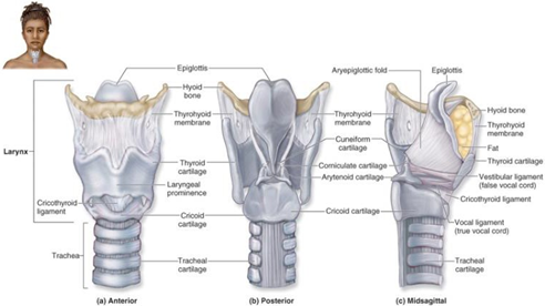
Picture 1. Anatomy larynx. 8
2 Definition
Squamous cell carcinoma (SCC) is a malignant tumor that shows squamous differentiation, such as intercellular bridges and/or keratinization. SCC originates from squamous epithelium or from respiratory epithelium that undergoes squamous metaplasia. 10
3 Epidemiology
Laryngeal cancer occurs more often in men than in women. However, the incidence is increasing in various countries, including in women, possibly due to the increasing smoking habits. 9 A total of 210,606 new cases of laryngeal cancer were diagnosed in 2017 (2.76 new cases per 100,000 population) worldwide, with a prevalence in the same year of 1.09 million cases (14.33 cases per 100,000 population), resulting in 126,471 deaths (1.66 per 100,000 population) and 3.28 million disability - adjusted life years (DALYs) or 0.13% of all DALYs. The incidence and prevalence have increased by 12.0% and 23.8% respectively over the past three decades, while mortality has decreased by about 5%. Among all types of cancer, laryngeal malignancies rank order 22nd in terms of incident (0.89% from all over type of cancer), 18th in matter prevalence (1.44% from all over type cancer), 18th in matter DALYs (1.50% from all type cancer) and 1.39% from all over death consequence cancer. 11
4 Risk Factors
There are many risk factors associated with laryngeal carcinoma, especially squamous cell carcinoma. These factors have been shown to play an important role in the development of carcinogenesis. Various agents are involved in the process. This, Which cause mutation And transformation development cell. The following is a brief overview of the major risk factors for laryngeal SCC. 9
Smoke
Tobacco smoking is the most significant risk factor for laryngeal cancer. Extensive research since the 1950s has demonstrated an association between tobacco smoke and the development of head and neck cancers. In the United States, approximately 95% of patients with laryngeal cancer are smokers. The carcinogenic effects of cigarette smoking on laryngeal cancer are now well established, but the specific risks for different types of smoking are less clear. 9
Alcohol
A recent systematic review by Levesque et al. 11 showed that dietary intake alcohol increase risk disease infectious And cancer, including laryngeal SCC, in a dose-dependent manner. In addition, several studies have speculated on the possible interaction between alcohol intake and genetic susceptibility, particularly in relation to genetic polymorphisms of the enzymes alcohol dehydrogenase (ADH) and aldehyde dehydrogenase (ALDH). 9
Opium
HPV
Opium is an illegal substance derived from the opium poppy plant that contains various alkaloids. The International Agency for Research on Cancer (IARC) has classify opium as agent carcinogenic to man If sucked or digested in various form like raw opium, opium waste, or opium resin. 12
Although smoke still become factor risk main cancer larynx, HPV infection is commonly found in nonsmokers and young patients with laryngeal cancer. Several subtypes of HPV have been identified, some of which show a stronger correlation with transformation. malignant mobile. In between subtype This, HPV-16 often found in laryngeal cancer specimens. 9
Many studies have shown that smoking can cause persistence of HPV infection. This is due to the relationship between poor oral hygiene and the risk of developing HPV. HPV positive in squamous cell carcinoma squamous. Dangerous type
of the HPVs most frequently found in squamous cell carcinoma cases is HPV-16 And HPV-18 Which can cause infection cornice, abnormality cells, and cancer. About 90% of cancers in the oropharynx are associated with HPV, especially type HPV-16 And type HPV-18 found more A little on head and neck cancer cases. It has been reported that patients with HPV-positive squamous cell carcinoma have a lower risk of death and recurrence due to responding well to chemotherapy and radiotherapy compared to HPV-negative patients. 9
Helicobacter pylori And GERD
Recent studies have shown that there seems to be a correlation between H. pylori infection and laryngeal carcinoma. However, further research with population patient Which more big And data Which more comprehensive studies are needed to confirm this relationship conclusively.9
Factor other
Other less studied risk factors that may be less involved in head and neck cancer include genetic susceptibility, certain occupations, exposure to air pollution, and previous exposure to radiation therapy. Certain genetic factors can contribute to vulnerability somebody to head and neck cancers, including laryngeal cancer. Genetic studies have identified certain gene variants that may increase risk, but more research is needed to fully understand the extent of their influence. Individuals who have had prior radiation therapy for head and neck cancer may have an increased risk of developing a second primary tumor, including laryngeal cancer. However, the risk is influenced by factors such as the radiation dose, the duration since treatment, and the individual's susceptibility. 9
5 Histopathology
SCC can be ulcerated, endophytic, flat, exophytic, polypoid, or verrucous, ranging from small mucosal thickenings to occlusive masses. The tumors are erythematous to brownish white, often firm. SCCs range in degree from squamous to Which varies related with invasion. SCC generally shared become 3 histological grade (well, moderately or poorly differentiated), with or without keratinization (see Figure 2.1). SCC shows a desmoplastic stromal reaction, often time invasion perineural or lymphovascular, And disturbance membrane basal by cord cells, nests, islands, or individuals. There is loss of polarity, disorganization, dyskeratosis, And formation pearl keratin. 13 Bridge between cell seen between cells Which own improvement ratio nucleus-cytoplasm, turbidity cytoplasm, and irregular polygonal shape. There is irregularity of nuclear chromatin, prominent eosinophilic nucleoli, and increased mitosis, including atypical forms. Keratinization Which develop with Good No seen on tumor differentiated bad (Look Picture 3). On tumor Which differentiate bad or high-grade tumors, immunohistochemical studies for CK5/6, p63, p40, and epithelial membrane antigen (EMA) can help confirm the epithelial nature of the proliferation. 13
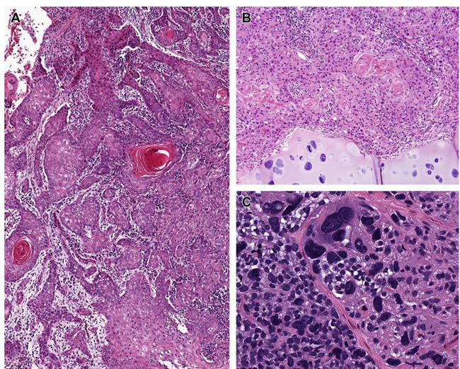
Figure 2. (A) Highly invasive, well-differentiated keratinizing SCC with keratin pearls. (B) Moderately differentiated keratinizing SCC showing cartilage invasion. (C) Poorly differentiated SCC with marked pleomorphism.
6 Diagnosis
Initial evaluation of patients with suspected laryngeal cancer is a detailed history. The history is done to find signs and symptoms of laryngeal carcinoma and to find any risk factors that the patient has. In addition, a complete physical examination of the head and neck should be performed, including examination of the mucosa of the upper aerodigestive tract, because tumors often occur primary multiple on patient with tumor head And neck. Recommended also to assess speech and swallowing function, communication needs, nutrition, health behavior and availability of social support. Pre-therapy dental evaluation is also recommended for patients who will undergo radiation due to the risk of damage, infection, and osteoradionecrosis due to treatment. If there is a suspicion of malignant tumor, this needs to be confirmed by biopsy of the lesion for histopathological study. Finally, radiological imaging is performed to see the exact anatomical details regarding the localization and extension of the tumor. 14
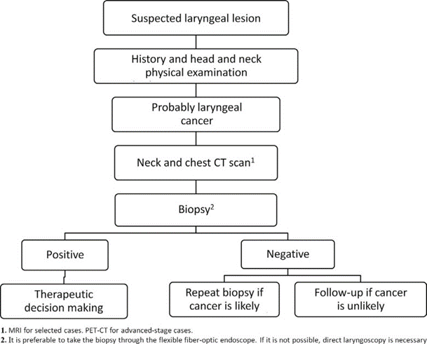
Picture 3. Algorithm diagnostic lesi larynx. 14
The staging of laryngeal carcinoma is determined by the location of the involvement of the larynx by the disease, thus requiring a thorough understanding of the anatomy of the larynx. The classification of the stage of laryngeal cancer is determined by the extent of involvement. subsite by tumor primary, mobility tape voice, expansion extranodal,
and the presence of metastasis. Tables 2.1 and 2.2 present the TNM staging system. from American Joint Committee on Cancer Which shared based on sublocation of the larynx followed by stage grouping. 15
Table 2.1 Category Tumor Primary (T)
Category | Supraglottic | Glottic | Subglottic |
T1 |
| Only true vocal cord involvement | Only subglottic involvement |
T1a | - | Ribbon involvement single voice | - |
T1b | - | Ribbon involvement bilateral sound | - |
T2 | Invasion of the supraglottis or adjacent glottis or areas outside the supraglottis without fixation larynx | Extension to the supraglottis or subglottis and/or impaired band mobility voice | Extensions to vocal cords |
T3 | Limited to the larynx withfixation tape sound and/or invasion of the postcricoid area, preepiglottic space, paraglottic space, or cortex part in thyroid | Limited to the larynx with invasion of the vocal cords and/or invasion of the paraglottic space and/or the cortex of the larynx. in thyroid cartilage | Extension into the larynx with fixation of the vocal cords and/or inner cortex involving the bones vulnerable thyroid |
T4a | Invasion throughouter cortex bone vulnerable thyroid | Invasion through outer cortex cartilage | Invasion through cricoid cartilage or thyroid |
| and/or invasion of tissue outside the larynx | thyroid and/or invasion networkin outside larynx | and/or invasion of external networks larynx |
T4b | Space invasion | Space invasion | Space invasion |
| prevertebral, | prevertebral, | prevertebral, |
| arterial sheath | wrapping | wrapping |
| carotid, or invasion | artery carotid, or | artery carotid, or |
| mediastinum structure | structural invasion | structural invasion |
|
| mediastinum | mediastinum |
Source: Patel SG, Lydiatt WM, Glastonbury CM, et al (2017)
Table 2.2 Category nodal regional (N)
Category | Involvement NodalRegional |
N0 | No There is Involvement Regional Nodal |
N1 | Gland SapClear Ipsilateral Single< 3 cm No There is Extensions Extranodal |
N2a | Gland SapClear Single, Ipsilateral, Between 3 cm And 6 cm,Without Extensions Extranodal |
N2b | Gland Sap Clear Multiple, Ipsilateral, No There is larger than 6 cm No There is Extensions Extranodal |
N2c | Gland SapClear Multiple, Bilateral/Contralateral None larger than 6 cm No There is Extensions Extranodal |
N3a | Every metastasis regional which are more big from 6 cm No There is Extensions Extranodal |
N3b | Every metastasis regional with Extranodal Extension |
Source: Patel SG, Lydiatt WM, Glastonbury CM, et al (2017)
7 Clinical manifestations
Patients with primary early stage tumors arising on the native vocal cords usually present with complaints of dysphonia and hoarseness, sore throat, odynophagia, dysphagia, called otalgia, painful local on cartilage thyroid, hemoptysis, obstruction road breath, stridor And adenopathy neck is characteristic features lesi glottis advanced stage. In general, persistent hoarseness (>15 days) in adult patients requires endoscopic visualization of the larynx to rule out tumor. Supraglottic tumors usually present with more advanced symptoms than glottic cancers when with symptom following: flavor No comfortable or sensation lump in throat (most common initial symptom), dysphagia, odynophagia, sensation of a lump in the throat, respiratory obstruction, hemoptysis, referred pain to the ipsilateral ear (via nerve vagus And auricular) or mass in neck; However, voice hoarse not a primary symptom of supraglottic cancer until the lesion becomes quite extensive. Primary subglottic tumors usually asymptomatic on stage beginning And No appear until the size is bigger carry on in where often happen dyspnea And stridor. Dysphonia relatively often occurs in advanced subglottic tumors due to true vocal cord involvement or direct extension to the recurrent laryngeal nerve. Late symptoms of laryngeal cancer include weight loss, dysphagia, bad breath and aspiration. 14
8 Inspection physique
A thorough clinical examination with rigid or flexible fiberoptic endoscopy is performed to assess the surface area of the primary tumor, vocal cord mobility. And do staging tumor beginning. Lesion big can expand beyond larynx to the adjacent base of the tongue, piriform sinus, or retrocricoid area and this should be assessed. Determination of vocal cord mobility also needs to be assessed. Vocal cord fixation of cancer larynx due to by invasion or damage muscle tape voice, invasion cricoarytenoid muscle or joint or recurrent laryngeal nerve invasion. Pain or tenderness on palpation or a small prominence over the thyroid cartilage suggests thyroid invasion. Endoscopy narrow band imaging (NBI) should used as Routine pharyngolarynx examination can help in early diagnosis of laryngeal tumors and monitoring after surgery For detection recurrence. Technique imaging other like Automated fluorescence, contact endoscopy, and optical coherence tomography (OCT) are increasingly used in ENT practice. Sometimes, laryngoscopy is required. direct with anesthesia general. Ventricle, area subglottis, apex The pyriform sinuses, and the retrocricoid area should be carefully examined. A detailed examination of the neck is mandatory whenever a laryngeal tumor is suspected. Regional metastases is factor prognostic negative Which most important, lower survival rate of 50%. 14
9 Imaging
If suspected existence malignancy, computed tomography (CT) scan and/or magnetic resonance imaging (MRI) with contrast should be performed to determine the extent of the submucosa and deeper tumor boundaries. CT is preferred because need time more short than MRI. Tumor mucosa small and superficial may not be detected on CT or MRI so endoscopy is mandatory before imaging studies. Axial diameter more than 10 mm, spherical shape, necrotic nodes of various sizes and nodes with indistinct mottled borders are generally accepted radiological criteria for diagnosing malignant nodes on CT and MRI. Common radiological criteria used for tumor involvement are asymmetric soft tissue protrusion or thickening, abnormal contrast enhancement, large mass, loss of normal fat planes and spaces, or a combination of these. 14
Tumors arising in the aryepiglottic fold appear as exophytic masses. or infiltrative. Tumor This expand folds aryepiglottic And spread into the paraglottic space, may also spread further anteriorly into the preepiglottic space or posteriorly to invade the pyriform sinus. Tumors originating from the false vocal cords are lateral masses with a strong predilection for submucosal spread into the paraglottic space. More extensive tumors may destroy the thyroid cartilage and spread transglottically into the glottis and subglottis. Distribution tumor to room paraglottic on CT or MRI seen as tumor tissue that replaces normal paraglottic fat. 14Glottic tumors usually originate from the anterior half of the vocal cords and spread to in commissure anterior. Abnormalities commissure front seen on CT or MRI as tissue thickening of more than 1 – 2 mm. Subglottic spread below the anterior commissure is seen as irregular thickening of the cricothyroid membrane. The tumor can gain access to extralaryngeal tissues through the membrane. cricothyroid. Cancer subglottis diagnosed when there is thickening anything between the airway and the cricoid ring. Due to its late presentation, invasion of the cricoid cartilage, trachea, and cervical esophagus with extralaryngeal spread is a common finding on radiological examination of these patients. In addition, the use of PET scan with 18F-fluorodeoxyglucose (FDG) combined with CT and PET allows for functional and anatomical evaluation. FDG examination has the potential to differentiate benign and malignant processes, tumor grade, identify metastases, and diagnose tumor recurrence. In the field of head and neck cancer, FDG is useful for detecting clinically occult recurrences and determining residuals after definitive radiotherapy. However, CT-PET is not useful for excluding metastasis in the neck with N0 because PET cannot detect very small tumors (<5-7 mm). 14
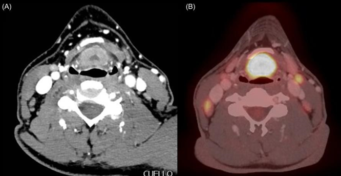
Figure 4. (A) Axial contrast CT shows a supraglottic tumor invading the pre-epiglottic cleft. (B) PET-CT shows a hypermetabolic focus of tumor active who occupies room pre-epiglottic in accordance with abnormality on CT scan. 14
10 Governance
Paradigm treatment moment This in management cancer larynx focus on healing while guard larynx when allow And strive for results Which give quality life best. For patient with cancer early stages (T1 and T2), endoscopic resection and open partial laryngectomy are attempted for complete resection while preserving the larynx. For patients with advanced disease (T3 and T4), treatment with chemoradiation therapy has emerged as the standard approach. Nevertheless, surgery plays an important role in the treatment of carefully selected patients. For those with advanced disease and poor function or those with contraindications to chemoradiation, surgery remains the mainstay of treatment. 16
Choose approach surgery versus non-surgical as treatment beginning for laryngeal cancer depends on individual patient factors such as age and comorbidities, location of the primary tumor, extent and volume of the primary tumor, and presence of metastases. gland sap clear or possibility metastasis in basin lymph nodes at risk of spreading, even without evidence of disease. Treatment options are also influenced by anterior commissure involvement and the ability to achieve adequate endoscopic visualization. The availability of surgical and radiation oncologists, as well as adequate rehabilitation services, are fundamental considerations. 16
Laryngeal cancer can be treated with a variety of surgical procedures. While Total Laryngectomy sacrifices the entire larynx, approaches in laryngeal conservation surgery aim to preserve the structure and function of the larynx while preserving the physiology of speech and swallowing without compromising the chances of cure. Laryngeal conservation surgery is performed in patients with early-stage laryngeal cancer with approaches including Transoral Laser Microsurgery and Open Partial Laryngectomy . Surgery larynx conservation must done only If expert surgery can confidently achieve tumor-free margins. Functional outcomes of laryngeal conservation surgery very depends on role surgery as the only one modality in treatment, because the use of postoperative adjuvant radiation therapy after incomplete resection may compromise functional outcomes, especially after open partial laryngectomy. 16,17
Modified Radical Neck Dissection
This procedure removes all the lymph nodes in the lateral neck (now known as level IV) and the spinal accessory nerve (CN XI), internal jugular vein (IJV), sternocleidomastoid muscle (SCM) and several structures. other in surrounding, including part caudal from gland parotid. Method This is indicated when the risk of cervical lymph node metastasis exceeds 15-20%. However, this procedure results in significant cosmetic deformity and loss of function. Over time, this procedure has been modified to reduce morbidity while maintaining oncologic efficacy. Modified Radical Neck Dissection (MRND) advocates preserving at least one of the important non-lymphatic structures (CNXI, IJV, or SCM). 18
For a neck dissection to be classified as an MRND, level IV must be spared, and at least one of the following structures must be spared: the spinal accessory nerve, the sternocleidomastoid muscle, and the internal jugular vein. Modified radical neck dissections are classified as type I, II, or III based on which structures are spared. In type I, MRND of CN XI is spared. On type II, MRND CN XI And IJV avoided. On type III, MRND CN XI, IJV and SCM were all spared. 19
Inoperable disease is an absolute contraindication to performing neck dissection. This includes invasion of the skull base or deep neck muscles. Carotid artery involvement may be considered an absolute contraindication. or relatively to surgery. If artery carotid Possible sacrificed, evaluation pre operation with studies occlusion balloon artery carotid with xenon CT or SPECT-CT imaging should be performed to determine the role of carotid reconstruction. No There is proof Certain Which show that action This increase survival, but is sometimes performed if the risk of carotid burst is felt to be high. 20
Total Laryngectomy
Total laryngectomy is a radical operation that consists of removing the entire larynx, including bone vulnerable thyroid And cricoid, bone hyoid And part of the thyroid gland. 13 The following are indications for Total Laryngectomy: 21
Advanced laryngeal tumor with cartilage destruction and anterior extra-laryngeal spread with a damaged larynx that is unlikely to function again even if anatomically preserved (laryngeal dysfunction, airway obstruction, or severe aspiration).
Circumferential submucous disease with or without bilateral vocal cord paralysis.
Involvement tumor joints cricoarytenoid bilateral or posterior commissure .
Involvement wide on bone thyroid prone .
Expansion subglottis with wide invasion on cartilage cricoid.
Tumor hypopharynx originate or spread to mucosa postcricoid.
Extensive resection of the pharynx or base of the tongue in patients at high risk for aspiration problems.
Tumor primary on cartilage cricoid or thyroid.
Necrosis radiation on larynx Which No get management medical.
Aspiration chronicle consequence incompetence glottis Which make patient not suitable candidates for conservative treatment.
Cancer thyroid stadium carry on with laryngeal invasion .
Although Total Laryngectomy often give opportunity best for healing, the consequences is a tracheostoma permanent and the disappearance original sound. Complications from modality This Which most general is infection wound And pharyngocutaneous fistula, happen on 50% patient Which previously accept radiation. Therefore That, flap network vascularized pedicle or free For cover Pharyngeal closure may be recommended to reduce the risk and severity of fistula. 16Various model incision in Total Laryngectomy explained in picture
2.1. A vertical midline incision provides direct access to the larynx but exposes exposure lateral Which limited to neck. Incision shaped T And Y double incision provides more exposure to the neck, but this incision involves a trifurcation, Which result in supply blood bad And results cosmetics Which poor. The Trap-door incision has been abandoned and replaced by contemporary single-stage reconstruction . The Apron incision is considered a better option. most liked For laryngectomy Because give results cosmetics best. While the extended apron incision is the main choice in patients who show tumors with lower extension or tumors that require extended apron neck dissection. 22
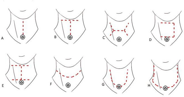
Picture 2.2 (A) Incision midline vertical , (B) incision T-shaped , (C) incision horizontal double-Y , (D) trap-door incision , (E) double trap-doors , (F) apron incision
(separate tracheostoma) , (G) incision apron (tracheostoma-incorporated) , and (H) extended apron incision 22
Fasciocutaneous Flap
Surgical flaps are usually classified based on their vascularization pattern and proximity to the primary defect. In terms of blood supply, flaps can be classified as random And axial . Flap random supplied by plexus subdermal skin And not supplied by vessels blood specific. On the contrary, the flap axial supplied by system arteriovenous certain. Flap axial usually considered more reliable than random flaps on the basis of increased perfusion in the distal part. 23
Based on the location of the defect with the location of the flap, it can be divided into local, regional, and free flaps. Local flaps are located adjacent to the location of the defect that occurs. Local flaps such as rotation flaps, advancement flaps , and transposition flaps. Durability life flap local depends on perfusion Which adequate. Flap regional and free flaps usually have axial-based vascularization, where they depend on a specific vascular pedicle for survival. Examples of regional flaps is flap pectoralis major myocutaneous, Which supplied by branch from the thoracoacromial artery. The use of free flaps requires microsurgical techniques. so that own limitations from aspect operator However own function and better aesthetics. 23
Faciocutaneous flap is a part of the axial flap whose tissue consists of from layer skin, network subcutaneous And fascia in. Flap faciocutaneous on the anterior chest under the clavicle bone or what is known as the deltopectoralis flap is often used for reconstruction in the head and face region by Because location Which near, texture skin And color skin. Flap deltopectoralis This flap receives its blood supply from the internal mammary perforator artery . This flap contains Lots supply artery And vein Which connected so that No causing necrosis of the flap. This faciocutaneous flap is also preferred compared to the myocutaneous flap because it is thinner and easier to use. Another advantage in using this faciocutaneous flap is that it does not require microsurgical tools and techniques so that the time required for surgery is shorter when compared to using a free flap so that it is more often chosen in patients with underlying comorbid diseases and patients with severe disease. Deltopectoralis faciocutaneous flap This Also own lack like range rotation Which limited and defects in the donor especially if skin grafting is required. 23
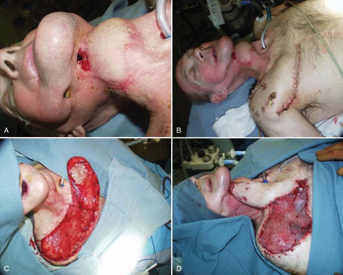
Figure 7. Deltopectoral flap. A, Hypopharyngeal defect. B, Delayed deltopectoral flap with split-thickness skin graft. C, Elevation of the deltopectoral flap with skin graft to demarcate the defect. D, Deltopectoral flap with skin graft placed over the donor defect. 23
Open Partial Laryngectomy
Open Partial Laryngectomy is a laryngeal conservation surgical technique that includes a variety of open surgical techniques, ranging from hemilaryngectomy to supracricoid partial laryngectomy. Complications of Open Partial Laryngectomy include bleeding, infection, laryngocutaneous fistula, poor wound healing, airway obstruction requiring tracheostomy, aspiration pneumonia, dysphagia, and dysphonia. Open Partial Laryngectomy is sometimes used in the primary treatment of T3 supraglottic cancers that cannot be performed with transoral laser microsurgery. In vertical partial laryngectomy, a vertical incision is made through the thyroid cartilage near the anterior commissure and just anterior to the posterior border cartilage thyroid, to resection tape voice true And subglottis closest, ventricles, false vocal cords, and aryepiglottic folds. Vertical partial laryngectomy when This only intended for for part small patient with tumor T1 small or small T2 involving the anterior commissure. Horizontal partial laryngectomy, or supraglottic laryngectomy, consists of resection of the entire part of the larynx that located in on glottis, including epiglottis And room preepiglottis, with while preserving the native vocal cords and arytenoids. Horizontal partial laryngectomy may be offered to select patients with tumors that extend beyond the traditional boundaries, which include T3 and T4 supraglottic tumors involving 1 vocal cord and 1 arytenoid and extending into the pyriform sinus or base of the tongue. 16
Transoral Laser Microsurgery
Transoral Laser Microsurgery (TLM) is a minimally invasive endoscopic approach that combines the use of a suspension laryngoscope with an operating microscope, a tissue-cutting laser, and microsurgical instruments to resect primary tumors. Carbon dioxide and Nd:YAG lasers produce beams with frequency light Which absorbed by water, so that minimize network damage. The concept of TLM is different from the open partial surgical approach. In Open Partial Laryngectomy , resection is performed in a systematic manner, removing certain anatomical structures according to a well-reported standard approach (eg: systematic resection of the thyroid cartilage in supracricoid laryngectomy), whereas in TLM, resection is based on the location and invasion of the tumor. The surgeon removes the abnormal tissue with its margins and preserves the normal structures to ensure adequate functional results. TLM is mainly indicated for early-stage vocal cord cancer (Tis, T1, and T2). 24
CONCLUSION
Based on description histology, Wrong One type carcinoma larynx Which The most common is squamous cell carcinoma (SCC). SCC is a malignant epithelial tumor that shows evidence of squamous differentiation and is a malignant neoplasm of the larynx. Which most important And most general. A number of factor risk has proven increase risk incident SCC Larynx Wrong the only one Which most main is smoking. Description clinical patient with carcinoma larynx depends on anatomical area of the larynx affected. Current management of laryngeal SCC consists of surgical and non-surgical procedures. Choosing a surgical versus non-surgical approach as a treatment beginning For cancer larynx depends on factor individual patient such as age and comorbidities, location of the primary tumor, area and volume of the primary tumor, and existence metastasis gland sap clear. A number of method surgery Which moment These are still applied are Modified Radical Neck Dissection, Total Laryngectomy, Fasciocutaneous Flap, Open Partial Laryngectomy, Transoral Laser Microsurgery.
REFERENCE
Cavaliere, M.; Bisogno, A.; Scarpa, A.; D'Urso, A.; Marra, P.; Colacurcio, V.; DeLuca, P.; Ralli, M.; Cassandro, E.; Cassandro, C. Biomarkers of laryngeal squamous cell carcinoma: A review. Ann. Diagn. Pathol. 2021, 54, 151787.
2. Ciolofan, MS; Vlăescu, AN; Mogoantă, CA; Ionit,ă, E.; Ionit,ă, I.; Căpitănescu, AN; Mitroi, MR; Anghelina, F. Clinical, histological and immunohistochemical evaluation of larynx Cancer. Curr. Health Sci. J. 2017, 43, 367–375.
3. Nocini, R.; Molteni, G.; Mattiuzzi, C.; Lippi, G. Updates on larynx cancer epidemiology. Chin. J Cancer Res. 2020, 32, 18–25.
4. Thompson LDR. Laryngeal Dysplasia, Squamous Cell Carcinoma, and Variants. Surg Pathol Clin. 2017 Mar;10(1):15–33.
5. Steuer CE, El-Deiry M, Parks JR, Higgins KA, Saba NF. An update on larynx cancer: An Update on Larynx Cancer. CA Cancer J Clin. 2017 Jan;67(1):31–50.
6. Obid R, Redlich M, Tomeh C. The Treatment of Laryngeal Cancer. Oral Maxillofac Surg Clin. 2019 Feb 1;31(1):1–11.
7. Koroulakis A, Agarwal M. Laryngeal Cancer. In: StatPearls [Internet]. Treasure Island (FL): StatPearls Publishing; 2022 [cited 2023 Feb 11]. Available from: http://www.ncbi.nlm.nih.gov/books/NBK526076/.
8. Thompson LDR, Wenig BM. Diagnostic Pathology: Head and Neck. Elsevier Health Sciences; 2018. 1193 p.
9. Liberale, C.; Soloperto, D.; Marchioni, A.; Monzani, D.; Sacchetto, L. Updates on Larynx Cancer: Risk Factors and Oncogenesis. Int. J. Mol. Sci. 2023, 24, 12913. https://doi.org/10.3390/ijms241612913
10. Zidar N, Gale N. Cancer of the Larynx; Pathology and Genetics. In: Reference Module in Biomedical Sciences [Internet]. Elsevier; 2018 [cited 2023 Feb 11].
11. GBD 2017 Disease and Injury Incidence and Prevalence Collaborators Global, regional, and national incidence, prevalence, and years lived with disability for
p. B9780128012383652000. Available from: https://linkinghub.elsevier.com/retrieve/pii/B9780128012383650924354 diseases and injuries for 195 countries and territories, 1990-2017: a systematic analysis for the Global Burden of Disease Study 2017. Lancet. 2018;392:1789–858. doi: 10.1016/S0140-6736(18)32279-7.
12 Levesque, C.; Sanger, N.; Edalati, H.; Sohi, I.; Shield, K.D.; Sherk, A.; Stockwell, T.; Butt, PR; Paradis, C. A systematic review of relative risks for the relationship between chronic alcohol use and the occurrence of disease. Alcoholism 2023, 47, 1238–1255.
13. Lester DR Thompson, MD. Laryngeal Dysplasia, Squamous Cell Carcinoma, and Variants. Surgical Pathology 10 (2017) 15–33
14. López Álvarez F, Rodrigo JP. Cancer of the Larynx: Diagnosis and Treatment. In: Reference Module in Biomedical Sciences [Internet]. Elsevier; 2018 [cited 2023 Feb 11]. p. B9780128012383654000. Available from: https://linkinghub.elsevier.com/retrieve/pii/B9780128012383653187
15. Patel SG, Lydiatt WM, Glastonbury CM, et al. Larynx. In: Amen MB, editor. AJCC cancer staging manual. 8th edition. New York: Springer; 2017. p. 149.
16 Randa Obid, BAa , Magi Redlich, BSNb , Chafeek Tomeh, MD, MPHc. The Treatment of Laryngeal Cancer. Oral Maxillofacial Surg Clin N Am 31 (2019) 1–11 https://doi.org/10.1016/j.coms.2018.09.001
17. Forastier A A, Ismail N, Lewin JS, et al. Use of larynx preservation strategies in the treatment of laryngeal cancer: American Society of Clinical Oncology clinical practice guideline update. J Clin Oncol 2018;36(11):1143–69.
18. Popescu B, Berteşteanu SV, Grigore R, Scăunaşu R, Popescu CR. Functional implications of radical neck dissection and the impact on the quality of life for patients with head and neck neoplasia. J MedLife. 2012 Dec 15;5(4):410-3.
19. Rodrigo JP, Grilli G, Shah JP, Medina JE, Robbins KT, Takes RP, Hamoir M, Kowalski LP, Suarez C, Lopez F, Quer M, The Bookkeeper CC, with Bree R, Coskun H, Rinaldo A, Silver CE, 20. Ferlito A. Selective neck dissection in surgically treated head and neck squamous cell carcinoma patients with a clinically positive neck: Systematic review. Eur J Surg Oncol. 2018 Apr;44(4):395-403
21 Bäck LJJ, Aro K, Tapiovaara L, Vikatmaa P, de Bree R, Fernández-Álvarez V, Kowalski LP, Nixon IJ, Rinaldo A, Rodrigo JP, Robbins KT, Silver CE, Snyderman CH, Suarez C, Takes RP, Ferlito A. Sacrifice and extracranial http://dx.doi.org/10.1016/j.path.2016.10.003. reconstruction of the common or internal carotid artery in advanced head and neck carcinoma: Review and meta-analysis. Head Neck. 2018 Jun;40(6):1305- 1320
22. University of Iowa Health Care [Internet]. Total Laryngectomy | Iowa Head and Neck Protocols. Iowa City: University of Iowa. 2019 [cited 2020 Dec 31]. Available from: https://medicine.uiowa.edu/iowaprotocols/total-laryngectomy
23. Chotipanich. A Total Laryngectomy: A Reviews of Surgical Techniques. Cureus 13(9): (September 22, 2021) e18181. DOI 10.7759/cureus.18181
24. McCarty JL, Corey AS, El-Deiry MW, Baddour HM, Cavazuti BM, Hudgins PA. Imaging of surgical free flaps in head and neck reconstruction. American Journal of Neuroradiology. 2018;40(1):5–13. doi:10.3174/ajnr.a5776
25. Hans, S.; Baudouin, R.; Circiu, MP; Couineau, F.; Oral, Q.; Crevier-Buchman, L.; Lechien, JR Laryngeal Cancer Surgery: History and Current Indications of Transoral Laser Microsurgery and Transoral Robotics Surgery. J. Clin. Med. 2022, 11
Comments