INSTALLATION PIPE GROMMET ON PATIENT OTITIS MEDIA EFFUSION
I Made Nudi Arthana
Otitis media effusion (OME) is a buildup of fluid in the middle ear. with membrane timpani Which Still intact without accompanied by with sign signs of infection acute. Otitis media effusion or often too referred to as otitis media Serous effusion will generally heal on its own within two to four weeks, but in some cases, it can last for months, even years. Chronic effusion in the ear often results in hearing loss. 1.3 OME often attacks children aged 1 to 3 years, followed by school age, namely 4 to 6 years. As many as 90% child age 10 year at least Once experience One time OME episodes. Although rare, otitis media with effusion can also affect adults. Cases of otitis media with effusion in adults usually occur after the patient has experienced infection channel breathing on Which heavy like sinusitis, reaction severe allergies, drastic changes in air pressure after flying or diving, or etiology other Which can cause dysfunction tube eustachius. Based on the report WHO regarding epidemiology otitis media, prevalence of otitis media effusion in Indonesia reached 3.8%. 3.4 Otitis media with effusion is clinically characterized by decreased function of the tympanic membrane and middle ear due to accumulation of fluid in the middle ear. The accumulation of fluid in the middle ear can be serous or mucoid and causes conductive hearing loss , ringing in the ears, a feeling of fullness in the ear, and sometimes can cause pain due to changes in pressure. 2,5,6
LITERATURE REVIEW
Anatomy Ear
On basically in a way anatomy ear shared become three parts big, namely the ears outside, ear middle, And ear in. Ear outside consists of from leaf ears and canal acoustician external. One third outside canal acoustician external composed of cartilage Which contain follicle hair And gland ceruminose while the inner two-thirds are bone parts covered by epithelium. The outer ear functions to capture vibration stimuli sound from outside towards the tympanic membrane. 11
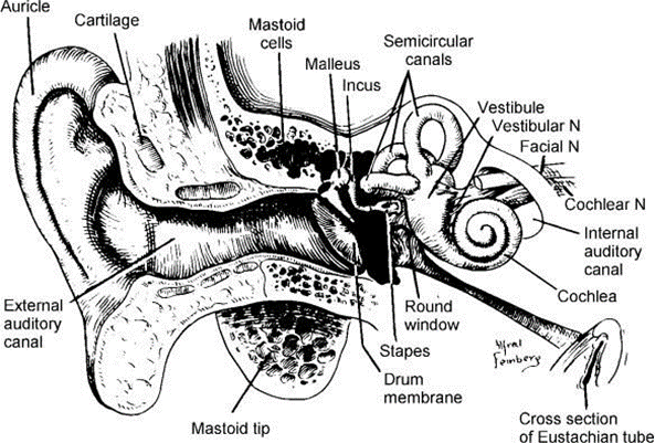
Picture 2.1. Anatomy ear 12
Anatomy Ear Outside
The outer ear consists of the auricle, ear canal and tympanic membrane. part lateral. Leaf the ear consists of from bone vulnerable elastin And The skin functions to collect sound waves, while the ear canal transmits them voice going to membrane timpani. Liang ear shaped S with a length of 2.5-3 cm. The outer third consists of cartilage containing many cerumen glands and hair, while the inner two-thirds consist of bone with little cerumen. 11
Anatomy Middle Ear
The middle ear is a cavity that contains air and keeps air pressure balanced. The middle ear is cube-shaped which functions to conduct sound sound from ear outside to ear in. Part front room The ear is limited by the tympanic membrane, while the inside is limited by the foramen ovale and foramen rotundum. In the middle ear space there are the following parts: 11
Membrane Timpani
Membrane timpani shared become two part, that is pars flaccida which is located at the top and pars tensa which is located at the bottom. Pars tensa is the part most big consists of from three layer. Layer outside called layer consists of from a smooth pseudostratified epithelial layer that normally reflects light. The inner layer called the mucosal layer, is a layer bordering with tympanic cavity and the layer between them. This layer consists of two layers of fibrous connective tissue that unite with a fibrocartilage ring that surrounds the tympanic membrane. The pars flaccida does not have a fibrous layer, so this is the first part to experience retraction when there is negative pressure in the ear. The tympanic membrane functions as a receiver of sound waves sound. Every There is wave sound Which enter hallway ear will be received by the tympanic membrane, then the tympanic membrane will bulge inward towards the middle ear and will touch the auditory ossicles, namely the malleus, incus and stapes. The auditory ossicles will transmit the sound waves to the inner ear . 11
The bones hearing
The auditory ossicles consist of the malleus, incus and stapes. The long process of the malleus is attached to the tympanic membrane, the malleus is attached to the incus, and the incus attached to stapes. Stapes located on window oval Which associated with cochlea. Third bone the to form series bone Which across the middle ear and merges with the tympanic membrane. 11
Cavity Timpani
The tympanic cavity is divided into three parts that are connected to the tympanic membrane plate, namely the epitympanum, mesotympanum and hypotympanum. The epitympanum is limited by a thin protrusion, namely tegmen tympani. The anterior part of the epitympanum contains the ampulla of the superior canal. In the anterior part of the ampulla of the superior canal there is the geniculate ganglion which is sign end front room Atik. Atik on part posterior narrows into the entrance to the mastoid antrum, namely the aditus ad antrum. Mesotympanum, in part medial restricted by capsule otic Which located more low than
n. Facialist pars tympani. The hypotympanum is a shallow space located lower than the tympanic membrane. The hypotympanum extends from the lateral semicircular canal to the posterior semicircular canal anteromedially. sine sigmoid. Corner This will found with throw away as clean as possible the mastoid pneumatization cells between the lateral semicircular canal and the sinodural angle. Trautman's triangle is the area located behind the antrum which restricted by the sigmoid sinus, lateral sinus And bone labyrinth. Limit medial is plate Dura posterior fossa. 11
Tube eustachian tube
Tube eustachian connects ear middle with nasopharynx. The length of the adult Eustachian tube varies between 31-38 mm. In infants and children the size is more short And more horizontal so that Secret from the nasopharynx easier to enter the middle ear. The anteromedial two-thirds tube (towards the nasopharynx) has a cartilage wall, while the rest (towards the tympanic cavity) has a bone wall. This cartilage wall is incomplete, the lower and lower lateral walls are connective tissue that joins the M. Tensor and levator velli palatini. The Eustachian tube will keep growing long and will forming a larger angle from the horizontal plane at the age of 5-7 years. 11

Figure 2.2. Anatomy membrane timpani 13
Anatomy Inner Ear
The inner ear consists of two parts, namely the bony labyrinth and the membranous labyrinth. The bony labyrinth consists of the cochlea, vestibule, and semicircular canals, while the membranous labyrinth consists of the utricle, saccule, cochlear duct, and semicircular ducts. The cavity of the bony labyrinth is lined by a thin layer of internal periosteum or endosteum, and is mostly filled with trabeculae (its arrangement resembles a sponge). The cochlea (snail house) is shaped like two and a half circles. End or peak cochlea called helicotrema, connect
perilymph scale vestibule (adjacent on) And scale timpani (adjacent lower). Between the scala vestibuli and scala tympani is the scala media (cochlear duct). The scala vestibuli and scala tympani contain perilymph with 139 mEq/l, while the scala media contains endolymph with 144 mEq/l. This is important for hearing. The base of the scala vestibuli is called the vestibular membrane (Reissner's Membrane) while base scale media is membrane basilar. On membrane This located the organ of Corti which contains organelles important for peripheral nervous mechanisms hearing. Organ Corti consists of from One line cell hair in Which containing
3,000 cells and three rows of cells outer hair containing 12,000 cells. Afferent nerve endings and efferent stick on end lower cell hair. On surface cells Hair has stereocilia attached to a sheath above it which tends to be flat, known as membrane tectoria. Membrane tectoria secreted And supported by a platform located medially called the limbus. 11,14

Picture 2.3. Anatomy labyrinth membranous 12
Anatomy Bone Mastoid
Behind the middle ear cavity is the mastoid antrum protrusion from bone temporalis And this mastoid cavity associated with ear middle through adit ads antrum. Cavity mastoid is a cavity shaped like a triangle with its apex pointing caudally. The mastoid antrum is 12-15mm long, 8-10mm high, and 6-8mm wide. 11,14

On the superior part or roof of the mastoid antrum is bordered by the tegmen tympani, wall medial And floor from cavity mastoid This restricted by pars petrous and mastoid part of the temporal bone, the lateral part of the mastoid cavity is bordered by squama part outside from pars mastoid, on part medial from mastoid cavity there is aditus ad antrum which connects the mastoid cavity with the middle ear. At the time of birth the thickness of the antrum wall is only 1-2mm but when adults the thickness can reach 9-10mm. As previously stated that the mastoid cavity is shaped like a triangle whose apex will point caudally, and this part is the mastoid process. 12
Cavity mastoid is mastoid water cell the biggest Which is at in the petrous part of the temporal bone. The mastoid air cells themselves have many sizes, from the small one to the big one. In the section above and front from The shape of the mastoid air cell in the temporal bone tends to be irregular and large and contains air, but if we look towards the bottom, the size of the mastoid air cell tends to be irregular and large and contains air. mastoid cell This will the more shrink, And even on part apex water mastoid cell Which is at there size very small or even rather contains bone marrow. 12
Vascularization, Flow Lymph And Innervation Ear
Artery auricularis posterior And branch auriculotemporal from artery The superficial temporal artery originates from the external carotid artery and supplies blood to the auricle and the cranial cavity. ear lateral. Branch auricularis deep from artery maxillary give
blood supply to the more medial aspect of the ear canal and the outer surface of the tympanic membrane. 11 The vascularization of the middle ear is served by 6 arteries, namely the anterior branch of the maxillary artery which serves the tympanic membrane, the stylomastoid branch a. auricularis posterior Which serve ear middle And cells mastoid airway, petrosal branch of the internal meningeal artery, superior tympanic branch of the internal meningeal artery, pterygoid branch and tympanic branch of the internal carotid artery. 11
The inner ear receives its vascular supply from the labyrinthine artery, which is a branch of the antero-inferior artery cerebellar. auricular vein posterior and superficial temporalis flowing hole ear. Veins auricularis posterior continued with the sigmoid sinus. The middle ear drains venous blood through the pterygoid venous plexus And sine petrosus superior. Flow blood vein ear in through
v. internal auditory, v. cochlear aqueduct and v. vestibular aqueduct. 12 Streams lymph from ear middle flow to retropharyngeal And gland parotid,
while the Eustachian tube lymph flows to the retropharyngeal group. 5 The auricle and external acoustic canal are innervated by the auricular temporal branch of the mandibular division of the trigeminal nerve, the greater auricular nerve from the cervical plexus, the auricular branch of the vagus nerve, namely the Arnold nerve, and branches of the facial nerve. And glossopharyngeus. Part lateral, medial And posterior ear innervated by nerves auricularis major. Nerve Arnold to innervate pars osseous hole lower ear, the cartilaginous part of the posterior superior ear canal and the corresponding segment of the membrane timpani And simba concha. Liang ear part posterior superioris innervated by branches of the facial nerve. The contribution of the trigeminal and glossopharyngeal nerves is not well described. 11,12
Nerves main ear middle served by plexus tympanicus And nerve of Jacobson, which receives its main supply from the glossopharyngeal n. The tympanic plexus provides innervation on surface medial membrane timpani, cavity tympani, mastoid air cells and bony part of the Eustachian tube. The chordatympanic nerve is a branch of the facial nerve that enters the middle ear through the posterior canaliculus and runs to the medial surface of the tympanic membrane between the arm of the malleus and the long process of the incus above the attachment of the tensortympanic tendon. The chorda tympani also supplies taste to the anterior two-thirds of the tongue and supplies secretomotor fibers to the submaxillary and sublingual salivary glands. 11,12
Otitis Effusion Media
Definition
Otitis media with effusion (OME) is a buildup of fluid in the middle ear with an intact tympanic membrane without any signs of infection. Acute. Otitis media with effusion or often known as nonsuppurative otitis media, mucinous otitis media, secretory otitis media, and mucoid otitis media ( glue ear ). Otitis media is a common condition that occurs in children aged 1 to 3 years, followed by school entry age, namely 4 years to 6 years. 13 As many as 90% child age 10 year at least Once experienced a single episode of OME. Many cases can resolve spontaneously, but 30% to 40% experience recurrence after 3 months and 10% case persists until 1 year. On person mature, Ome not enough common, but Still causing significant morbidity. The average incidence of OME is 14% - 62%, but several other studies report lower average prevalence rates. OME is 2% - 52%. Kamaludin, stated that the incidence of otitis media with effusion in Indonesia is 3.9-6.9%. 14 Otitis media that occurs in the pediatric population, which is associated with many factors, including adenoid hypertrophy, upper respiratory tract infection, cleft palate, and exposure to cigarette smoke. OME in adults is less common but still causes considerable morbidity. Mills and Hathorn mentioned several etiologies of OME in adults including local malignancy, sinonasal disease, gastroesophageal reflux disease, tubal dysfunction, smoking, and HIV. Roozbahany et al studied the causes of OME in adults aged 18-65 years found 41.9% allergies, 35.4% reflux gastroesophageal (RGE), 45.2% of nasopharyngeal mass, and almost 9% of neoplasms. Chronic OME is defined as OME that persists for 3 months or more on examination or tympanometry. 13,14,15 Most patients with OME do not have tympanic membrane retraction and only a few chronic OME have develop become atelectasis. When happen atelectasis membrane timpani will experience retraction to the promontory and the auditory ossicles in the middle ear, which occurs obliteration part or all middle ear cavity, and the mucosa lining it ear still in the middle intact. Atelectasis ear middle can be reversible with the installation of a ventilation tube. Sadé found that the installation of a ventilation tube can improve the atelectasis. 27

Picture 2.5. Classification atelectasis 14
Pathophysiology
Pathophysiology Ome nature multifactorial between other infection virus or bacteria, Eustachian tube dysfunction, immunological status, allergies, environmental and social factors.1 16 There is a number of theory Which be the cause the occurrence disease otitis media with effusion, The first is Eustachian tube dysfunction. The Eustachian tube has three functions, namely regulatory function, protection function, and secretion. Conditions that can causes tubal dysfunction classified into two, namely functional factors and mechanics. Mechanical factors are caused pressure enlarged adenoids or neoplasm, process inflammation Which caused by infection, allergies, or trauma. Disruption of the tube function causes the aeration mechanism to the middle ear cavity. disturbed, drainage from cavity ear middle to cavity nasopharynx disturbed And disturbance mechanism protection cavity ear middle against reflux from the cavity nasopharynx. Consequence the disturbance cavity ear the middle will experience negative pressure. Negative pressure in middle ear causes an increase permeability capillary And furthermore happen transudation. Besides there is infiltration of the inflammatory cell population and glandular secretions. As a result of the accumulation Secret in middle ear cavity. Inflammation chronic in the middle ear will cause formation network granulation, fibrosis, And destruction bones. 14,15,16 Theory both of which cause occurrence of otitis media with effusion is there is a condition And syndrome Which influence form one third middle face and the base of the throat such as Down syndrome and cleft lip. Children with lips chippy can experience insertion tensor veli palatine Which abnormal in the sky - sky soft Which cause inability For open tube eustachius adequately during swallowing and opening the mouth. This condition greatly influences the increased risk of otitis media with effusion. 15,16
The third theory that causes otitis media with effusion is as a residual symptom of acute otitis media, where in children with acute otitis media, 45% were found to suffer from Otitis media with effusion in 1 month after that and 10% on 3 months after that. Things This Can happen estimated that pepsin on 60% of middle ear effusions cause upregulation of mucin genes which leads to increased secretion season Which Can become place develop breed for upper respiratory tract bacteria. 16
The fourth theory that causes OME is secondary and subclinical bacterial infections such as pepsin which increases secretion season, cytokines, and also positive bacteria found in the examination of Otitis media effusion. The last theory that also causes the disease otitis media with effusion is related to gastroesophageal reflux disease where the pepsin in the middle ear effusion comes from reflux 15,16
Factor immunological And factor allergy Also play a role in the occurrence O.M. In immunological factors that play a significant role in OME is secretory Ig A. This immunoglobulin is produced by glands in the mucosa of the tympanic cavity. Secretory Ig A is mainly found in mucoid effusions and is known as an immunoglobulin. Which active Work on the surface mucosa respiratory. His work namely blocking germs so as not to have direct contact with surface epithelium, by forming complex bonds. Direct contact with the epithelial cell wall is the first stage of germ penetration for tissue infection. Thus Ig A actively prevents infection germs. In allergic factors, the rationale used is the embryogenic analogy, where the tympanic mucosa originates from the same as the nasal mucosa. At least allergic manifestations in the Eustachian tube are the cause of chronic occlusion and subsequently cause effusion. However, from study level Ig E which become criteria allergy atopic, good the level in effusion and in serum does not fully support allergies as Cause. Etiology and the pathogenesis of otitis media due to allergies may be caused by one or more from mechanism like mucosa ear middle as organ target organ, swelling due to inflammatory processes in the Eustachian tube mucosa, nasal obstruction due to inflammatory processes, and aspiration of nasopharyngeal bacteria found in allergic secretions into the middle ear space 15,16,17
Diagnosis
Diagnosis of otitis media with effusion is not easy and there are significant differences according to the clinician's skills. To establish the diagnosis of OME, anamnesis, otoscopy, tympanometry, and audiometry are carried out. 19,20
Anamnesis
OME often occurs in children, although this disease rarely causes complaints. The history of this disease is usually known from the parents or teachers of the children. The complaints conveyed are an attitude of indifference towards voice surrounding, disturbance or delay talk, And Also learning achievement child Which the more decrease. On children Which more big or Adults usually feel an uncomfortable feeling in the ear (feeling full), ringing in the ears Which echo on ear, Possible Also obtained tinnitus. Anamnesis not in get complaint presence of signs of infection. 15,16 The medical history needs to be explored completely and the risk factors for OME in patients should be sought. Immunological status, nutritional status, history of allergies in patients and their families, and abnormalities congenital that accompanies, for example palatoschisis is factors that should be considered in patients suspected of having OME. 16,19
Otoscopy
Examination conducted to assess the condition, color, and translucency of the tympanic membrane. Various changes due to abnormalities that occur in the tympanic membrane can be seen as follows 19,20 :
A dull, yellowish tympanic membrane changes the picture. translucent light besides That location triangle reflex light on 6 the anteroinferior quadrant is shortened, there may also be an increase in capillary blood vessels in the tympanic membrane. In cases with mucoid or mucopurulent fluid, the tympanic membrane is lighter in color.
Tympanic membrane retraction namely when the manubrium of the malleus appears shorter and more horizontal, the membrane appears more concave and the light reflex shortens. The color may turn slightly yellowish.
Atelectasis membrane timpani usually thinning, atrophy And Possible stick to incus, stapes And promontory, specifically on cases Which already continued, usually case This Because dysfunction tube eustachian tube And Ome Which Already walk in time that long.
Membrane timpani with cicatrix, gloomy until retraction heavy accompanied by with the atrophic part found in adhesive otitis media due to the occurrence of middle ear fibrotic tissue as a result of a previous inflammatory process that lasted a long time. This condition can be a complication of supurative otitis media or non-suppurative OME which causes damage to the middle ear mucosa. During healing, fibrotic tissue is formed which causes adhesions.
Fluid level water image or bubbles are usually found in OME with serous fluid.
A dark blue or purple tympanic membrane is seen in cases of hemotympanum. Which due to by fracture bone temporal, leukemia, vascular tumor of the middle ear. The lighter blue color may be due to barotrauma.
Otoscopic examination shows suspicion of OME if the following 19 signs are found :
No existence signs inflammation I
There is change color membrane timpani consequence reflex from existence fluid in the tympanic cavity
Membrane timpani looks more prominent
Membrane retraction or atelectasis
Got it water fluid levels or bubbles
Mobility membrane reduce or fixation
Sadness And Berco describe 4 stadium from retraction membrane timpani 27 :
stadium I : membrane timpani retraction light
stadium II : membrane timpani retraction And contact with incus or stapes
stadium III : atelectasis ear middle ( membrane timpani touch
promontory)
stadium IV : otitis media adhesive (membran timpani attached to the promontory)

Picture 2.6. Stadium retraction membrane timpani 14
Tympanometry
A tympanometer is a tool for determining the condition of the eardrum. from system middle ear. This measurement provides an overview of the mobility of the tympanic membrane, the condition of the auditory ossicle joints, the condition of the middle ear including the air pressure in it, so it is useful in identifying conduction disorders. and function tube Eustachian. 15 Results graph measurement tympanometry or tympanogram can be used to determine the picture of abnormalities in the middle ear. Although many variations of tympanogram forms have been found, in principle there are only three types, namely type A, type B, type C. Type A is a picture of tympanogram with the peak that sharp or steep Where pressure in middle ear range between 0 until -100 mm H2O. type This there is on ear normal middle ear, fixation, cicatrix, or ossicular discontinuity. Type B is relatively flat, indicating limited tympanic membrane movement due to fluid or adhesions in the tympanic cavity. A very flat graph may occur due to tympanic membrane perforation or excessive cerumen in the external ear canal. Type C tympanogram when the peak of the curve shifts at a pressure negative under -100 mm H2O, condition This happen on pressure negative in the tympanic cavity as a result of Eustachian tube function insufficiency. 19

Picture 2.7. Tympanogram 19
Audiometry
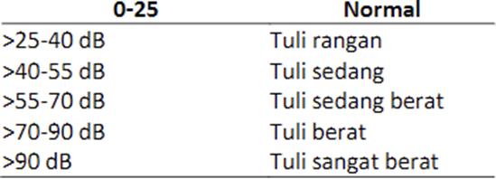
Audiometry is an examination to determine a person's hearing threshold. What is examined in an audiometry examination is the conduction hearing threshold. air conditioning (AC) and delivery bone (BC). On interpretation The audiogram must be written in which ear, the type of deafness, and the degree of deafness. determine degree of deafness, which counted only threshold just listen to the airflow.
Table 1. Degrees Deafness according to International Standard Organization 15
Hearing loss is more common in OME patients with fluid. Which thick (glue ears). Although thus a number of studies say there is no significant difference between serous and thick fluids on hearing loss, whereas volume fluid Which found on ear middle is more influential. Patients with OME on audiogram are often found to have hearing loss with mild conductive hearing loss (CHL). until currently so that No so affected with life a day-
days. Patients with severe conductive hearing loss may have had fixation or rupture of the ossicular chain . 22 The OME guidelines jointly developed by the AAFP and AAOHNS and AAP state that audiometry is one component of the examination of patients with OME. Audiometry is recommended on patient with Ome in 3 month or more or suspected to exist decline hearing meaningful. Based on statement in accordance with some studies, conductive deafness is often associated with OME and has an effect on process hear second ear, localization voice, perception talk in noise. 22,23
Radiology
Effective radiological examinations for OME screening include plain mastoid radiographs, which will but this examination has long been abandoned. CT-scan is very sensitive For to uphold diagnosis especially For get rid of the presence of complications of otitis media such as mastoiditis, sigmoid sinus thrombosis, or cholesteatoma, but this examination is not really necessary in establishing the diagnosis of OME. CT-scan plays an important role in unilateral OME patients to rule out the possibility of a mass in the nasopharynx. 22
Management
Generally OME resolves spontaneously within 3 months, conservative treatment in the form of steroid intranasal or steroid systemic, antibiotics, antihistamines, and decongestants are no longer recommended. So observation is carried out or watchful waiting for 3 months from the onset of effusion or since the diagnosis was made especially for children who are not at risk for speech and language disorders or learning disorders. Clinical practice guidelines from the American Academy of Otolaryngology-Head and Neck Surgery do not recommend the use of oral or intranasal corticosteroids. 27 This is also similar to Berkman et al. oral steroid administration during 2 months with results There is no benefit in using oral steroids as therapy for OME. Principi et al. said that giving decongestants is not recommended as a treatment for OME because it has not been proven to be effective and can increase risk of developing rhinitis medicamentosa. Treatment And control to allergy can reduce or cure OME. 19,20 Barazi et al said Desloratadine is effective in treating children with OME especially with a history of allergies, because it can accelerate the resolution of effusion and hearing loss, reducing the number of ventilation tube requirements. 10,24
Operative treatment is performed in cases where conservative treatment for more than 3 months does not heal. There are several indications for operative action in OME in the form of hearing loss associated with OME, suspicion of speech and language delay, autism spectrum disorder and other developmental disorders, Down syndrome or craniofacial disorder . To provide good results for drainage, myringotomy and installation of a tympanostomy tube or ventilation pipe are performed. 24,25 The tympanostomy tube was first introduced by Beverley Armstrong in 1954 and is still used until now. This tube designed to remain within the tympanic membrane and prevent rapid closure of the myringotomy incision. These tympanostomy tubes are small and well tolerated by the tympanic membrane. Tympanostomy tubes can be maintained for 1-2 years depending on the type used. Tympanostomy tubes must be placed in a normal area. Areas that are myringosclerotic will result in tube tympanostomy more easy let go And No well fixed. Tympanostomy tubes placed in normal areas will be surrounded by sclerotic tissue that allows the tympanostomy tube to be installed longer. Tympanostomy tubes are placed in the antero inferior quadrant area or poster inferior. Tube tympanostomy will maintained until patent tube function. 26,27 The purpose of installing a tympanostomy tube or ventilation tube is to eliminate fluid on ear middle, overcome disturbance hearing that occurs, preventing recurrence, preventing cognitive, speech development disorders, Language And psychosocial. There is Lots type tube tympanostomy, start from the shape, size, and basic materials used to make it. Based on size and shape, there are Which installed only For a number of month And There is Which maintained until some year. For example tube bobbin type installed For 6 until 18 month. This tube is removed when the tympanic membrane heals. While the T-type tube is installed for several years to prevent repeated installation. However, the disadvantage tube T-type This is cause perforation permanent And triggerthe occurrence of cholesteatoma. As with short-term tubes, long-term tubes also often become blocked by cell debris, requiring tube replacement. 26,27,28 Examples of tympanostomy tubes like Paparella, Shepard, Armstrong Gommet, Reuters Bobbin, and T tube.
Table 2. Type, size, And term usage time 26


Picture 2.8. Example tube tympanostomy 26
4 Complications
The further consequences of OME can result in hearing loss which will affect speech and intellectual development. Changes that occur in the middle ear can cause the disease to progress to atelectasis and adhesive otitis media. 21,23
Atelectasis and adhesive otitis media usually occur together with OME, although OME can resolve allowing aeration of the attic and mastoid, but not all patients with chronic OME develop atelectasis. Permanent damage due to OME can cause partial or total hearing loss. 23
There are two complications from installing a tympanostomy tube, namely intraoperative and postoperative. Complications intraoperative in the form of occurrence laceration, bleeding, or hematoma moment incision done, paralysis nerve facial Which happen moment injection of local anesthesia, incisions that are too wide and deep will cause bleeding in the mucosa, and tympanostomy tubes that fall into the middle ear. While post-operative complications can be purulent or mucoid otorrhea after done action, otalgia, debris or earwax which blocks the tympanostomy tube, cholesteatoma due to epithelial migration into the middle ear, natural detachment of the tympanostomy tube. 27
REFERENCE
Diaz RC, Kamal SM, Brodie HA. Middle Ear and Temporal Bone Trauma. In: Bailey BJ, editor. Bailey's Head and Neck Surgery-otolaryngology. Fifth Edition ed. Philadelphia: Lippincott Williams & Wilkins; 2014.
Wallace IF, Berkman ND, Lohr KN, et al. Surgical treatments for otitis media with effusion: a systematic review. Pediatrics. 2014:peds. 2013- 3228.
Berkman ND, Wallace IF, Steiner MJ, et al. Otitis media with effusion: comparative effectiveness of treatments. 2013.
Lin HP, Cheng PW, Tsao CH, et al. Bacterial role for post-irradiation otitis media with effusion in nasopharyngeal carcinoma. The Journal of International Advanced Otology. 2011;7(2):244-9..
Hsin CH, Chen TH, Liang KL, et al. Postirradiation otitis media with effusion in nasopharyngeal carcinoma patients treated by intensity‐modulated radiotherapy. The Laryngoscope. 2013;123(9):2148-53
Parhusip TD, Utomo BSR, Marlene L., Poluan FH, Falorin J., Nurfachri A., et a. Bacteria Reason Otitis Media in House Sick General Christian University of Indonesia. UKI Medical College. 2021;36(1).
Kamaludin D, Boesoirie TS, Soeseno B, Bambang P. The Effect of Using Nasogastric Tubes on the Incidence of Otitis Media Effusion.MKB.2011;43(1):42-48.
RosenfeldRM,ShinJJ,SchwartzSR,CogginsR,GagnonL,HackellJM,Hoeltin gD,HunterLL,KummerAW,Payne SC, Poe DS, Veling M, Vila PM,WalshSA,CorriganMD.ClinicalPracticeGuideline:OtitisMediawithEffu sion(Update). Otolaryngol Head Neck Surg.2016.
Shinta Dewi B, Christy P, Sagia Alsa N, Himayani R. Otitis Media with Effusion : Etiology, Pathophysiology, Pathogenesis, Epidemiology, Diagnosis, Management, Complications. Medulla. May 2023.
Farhat. Textbook of Ear Nose Throat Diseases and Head Neck Surgery. Medan:University of North Sumatra. 2019
S.ARennathaA,Artono.PerasnCytokines in Otitis Media with Effusion. 2017;10(1):37-44.
Doner F , Yariktas M, Damires M. The role of allergy in recurrent otitis media with effusion. J Invest Allergol Clin Immunol. 2014;14(4): 154-58.
Sanli A, Tasdemir O, Yilmaz SH. Prevalence of otitis media with effusion among primary school age children and etiopathogenic examination. Assoc of Otolaryngologists of India. 2011.
American Academy of Family Physicians, American Academy of Otolaryngology-Head and Neck Surgery and American Academy of Pediatrics 19 Subcommittee on Otitis Media with Effusion. Otitis media with effusion. Pediatrics 2004; 113(5): 1-18.
Robb PJ, Williamson I. Otitis media effusion. In: Watkinson JC, Clarke RW, editors. Scott- Brown's otorhinolaryngology head and neck surgery. 2. 8 ed. Boca Raton: Taylor and Francis Group; 2018. p. 115-36.
Banerjee A. Acute otitis media with effusion in adults. In: Watkinson JC, Clarke RW, editors. Scott-Brown's otorhinolaryngology head and neck surgery. 8. Boca Raton: Taylor & Francis Group; 2018. p. 971-5.
Rimmer J, Giddings CE, Weir N. History of Myringotomy and Grommets. J Laryngol Otol. 2007;121(10):911-6
Dhingra PL, Dhingra S, Disorders of middle ear. In : Disease of Ear, Nose, and Throat 7 ed. New Dehli: Elsevier India; 2017, pp. 69-71
Roderick P Venekamp, Martin Burton, Alice van Heijden. Antibiotics for Otitis Media with Effusion in Children. Cochrane Library, June 2016
Griffin G, Flynn CA, Bailey RE, Schultz JK. Antihistamines and/or decongestants for otitis media with effusion (OME) in children. Cochrane Database of Systematic Reviews. 2006
Cenggel S, Akyol MU. The role of topical nasal steroids in the treatment of children with otitis media with effusion and/or adenoid hypertrophy. International journal of pediatric otorhinolaryngology. 2006 Apr 1;70(4):639-45.
Lack G, Caulfield H, Penagos M. The link between otitis media and effusion and allergy: a potential role for intranasal corticosteroids. Pediatric allergy and immunology. 2011 May;22(3):258-66.
Szefler SJ. Pharmacokinetics of intranasal corticosteroids. Journal of allergy and clinical immunology. 2001 Jul 1;108(1):S26-31.
Berlucchi M, Salsi D, Valetti L, Parrinello G, Nicolas P. The role of mometasone furoate aqueous nasal spray in the treatment of adenoidal hypertrophy in the pediatric age group: preliminary results of a prospective, randomized study. Pediatrics. 2007 Jun 1;119(6):e1392-7.
Parish AB, All R, Hawk L, Pringles MI, Darby Y, Scattering G. Treatment of allergic rhinitis and its impact in children with chronic otitis media with effusion. Journal of Audiological Medicine. 2000 Jun 1;9(2).
Roland NJ, McRae RD, McCombe AW. Chronic suppurative otitis media. Key topics in Otolaryngology and head and neck surgery. 2001;2:38-41.
Sadness J, Berco E. Atelectasis and secretarial otitis media. Annals of Otology, Rhinology & Laryngology. 1976 Mar;85(2_suppl):66-72.
Comments