PREAURICULAR FISTULA
I Made Nudi Arthana
Preauricular fistula is abnormality congenital in the preauricular soft tissue, which is characterized by the formation of a fistula in the area. Usually there is hole small or pit on front helix ascending which is located in the anterior part of the outer ear.
LITERATUR REVIEW
Definition
Van Heusinger described the preauricular fistula for the first time in 1864. 2,5,8 Preauricular sinus or what is often called preauricular fistula is abnormality congenital benign on network soft preauricular and located on margin front from helix. 1.9 Fistula preauricular can be a simple hole or have a sinus tract and/or cystic components. 10 Similar terminology used interchangeably in the literature includes preauricular hole, fistula, tract, and cyst. 4
In general clinical, condition in where fistula preauricular become repetitive, periauricular granuloma occurs, or scars form due to repeated infections after fistulectomy, called as fistula preauricular complex. 7 After fistula infection occurs, cysts and fistulas need to be completely removed to control the infection and prevent complications. 2 The disease is most often seen on routine ear, nose, and throat examination, but can present as an infected, draining sinus. Preauricular sinus is more often unilateral but bilateral forms are sometimes inherited. The right side is more often involved and women are more likely than men. 11
Anatomy and Embryology
Accessory auricles (AA) or accessory auricles are abnormal growths from network Which remaining after formation leaf ear front at the stage beginning development embryo. Leaf ear outside develop from arch the first and second pharynx during the 4th week of pregnancy. The first pharyngeal arch produces three bulge small Which join to form structure leaf anterior ear. This prominence is initially located on the side of the mouth and then moves laterally and backward during development. Failure of the three prominences to fuse small on arch pharynx First believed become reason development
AA locations can occur anywhere along the migratory pathway within the triangle extending from the commissural groove. mouth to earlobe anterior, between helical spine and ear lobe. 12
Some studies describe this extra skin growth as tags preauricular. However, complement skin This can found in area buccal, intra-auricular, or precrural , all of which are distinct from the preauricular area. This Also can located in area prelobal, crural, or global, but No There is located in area tragus. 12 Fistula preauricular estimated originate from the fusion that Wrong from six bulge hearing His on Sunday sixth pregnancy. 5,10 This small protrusion is located at the junction of the first and second branchial arches, and branchial cleft anomalies may be associated. 10 Preauricular fistula occurs due to failure in process merger tubercle first And second from branchial arches. 2,6,9 This abnormality occurs due to failure of fusion of the tubercles on each of the first and second branchial arches that form the auricle during embryonic growth. 6,9
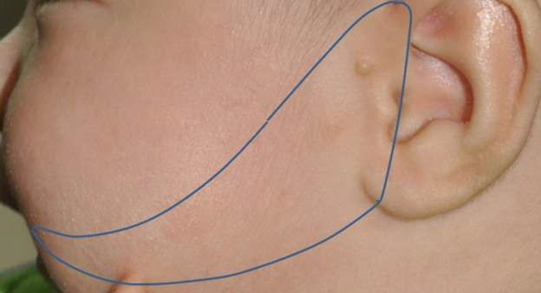
Picture 1 . A A in triangle arch. A A found in triangle mearkus from the groove of the oral commissure to the anterior auricle between the helical spine and the ear lobe. 12
Preauricular fistula is a congenital abnormality characterized by a small hole or foramen, usually located in part middle helix auricular. Seldom once a structure with similar characteristics and clinical course can be found in the pinna, tragus, or even in the postauricular area. 13 The structure can appear unilateral or bilateral, with possibility more from One fistula appears in one ear, especially on the earlobe. 6,13 Location of the opening of the preauricular fistula Which most general is edge front helix Which up, followed by the eminence of the cymba conchae. Furthermore, these openings are more often unilateral than bilateral. 8 The bilateral form is usually inherited by an incomplete autosomal dominant genetic trait. 6,
This structure is a simple epithelial invagination into the auricular stroma that can often become inflamed, causing leakage of fluid. from hole surface And create discomfort. The location which is close to the facial nerve can cause serious complications such as paralysis. face. History clinical Which thorough required For differentiate between a fistulized abscess and an inflamed preauricular fistula. In addition, chronic inflammation is also a risk factor for cancer formation, which can leading on development carcinoma cell squamous from fistula preauricular in the future. Therefore, interest in this structure is high among otolaryngologists, surgical specialists, and pediatricians. 13
A retrospective study using an extended imaginary tragus line in a way vertical For limit And categorize sine into the classic type (preauricular sinus) and the variant type (postauricular type). These variant types are then divided into type 1 (in the middle crus), type 2 (in the superior part of the crus), and type 3 (in the cymba concha), based on the location of the opening. Although abnormality This happen during process embryology, etiopathogenesis still not yet fully revealed. Moment This, there is three theory Which submitted: fusion incomplete from bulge arch branchial First, isolation folds cell ectodermal during formation leaf ear, And closing Which No perfect on part most dorsal of the first branchial cleft. 4
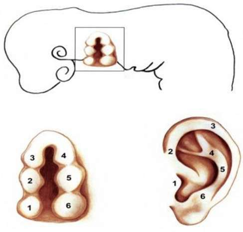
Picture 2 . Stage development ear. (1) Tragus (2) Crucible helix (3) Helix (4)
Antihelix (5) Antitragus (6) Lobules. 5
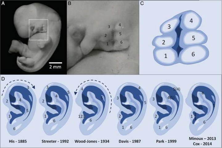
Figure 3. Theories proposed regarding the embryological development of the human auricle. (A) Looks lateral embryo original on specimen Carnegie stadium 17 (42– 44 day) 6521. (B) Close-up of the auricle process. (C) Simplified version of the auricle process. Numbers 1 to 3 have been described as the origin of the first pharyngeal arch and 4 to 6 as the derivatives of the second pharyngeal arch. (D) Proposed theory of the embryological development of the human auricle as described by several authors. The processes are represented by numbers 1 to 6. 14
Etiology
Preauricular fistula is caused by incomplete fusion of the six auditory crests or a defect during preauricular development. 8 Preauricular fistula can be sporadic or inherited, and has been mapped to a specific chromosomal location. 8,11 More than 50% of cases are unilateral and are most often sporadic. Bilateral cases are more likely to be inherited by incomplete autosomal dominance with reduced penetrance (nearly 85%). 5,11
The researcher in China has map possibility locus For congenital preauricular fistula on chromosome 8q11.1-q13.3 through family relationship analysis Which consists of from member Which caught impact And No caught impact. Preauricular fistula has been described as part of a number of syndromes and in association with renal or inner ear abnormalities. 11
Diagnosis
Anamnesis And Inspection Physique
A thorough history and head and neck examination are mandatory in all cases, looking for evidence of associated anomalies. 11 Most preauricular fistulas No show symptom And No need treatment. However, in the post-auricular type, surgical excision is recommended because the possibility of recurrent infection is quite high. 11,15 Only about 25% of preauricular fistula patients experience symptoms. 8 Asymptomatic conditions in this disorder sometimes cause patients to be unaware of their condition, causing patients to come to health services when obstruction and infection have occurred. 1,11 Infection conditions that are not treated quickly and properly can cause abscesses or secretions where if this condition is left untreated can trigger chronic infection and sepsis. 1
Sine preauricular is findings Which seldom happen And most often appears as a small hole near the anterior margin of the ascending portion of the helix. The opening Also has reported throughout margin poster superior helix, tragus or lobule. Preauricular sinuses may cause the formation of subcutaneous cysts that are closely related to the cartilage of the tragus and the anterior crus of the helix. 11 The most prominent clinical manifestation in affected individuals is a congenital abnormality of the external ear. Preauricular fistulas usually do not cause symptom Which real. However, a number of patient Possible come with local manifestations, such as erythema, swelling, pain, discharge of pus, swelling of the surrounding soft tissue, and skin damage due to infection. The symptoms This can impact Serious on cleanliness face And quality life as a whole. 3
Inspection Support
Preauricular fistulas are generally narrow, of variable length, but their orifices are usually short and small, protected and branch around the ear. outside. Long channel fistula varies between 3 until 22 mm. Methylene blue can used For to browse channel those. 5 Journey sine on subcutaneous tissue preauricular No constant. Hole Which seen Possible represent overall deformity, or marked sinus tracts of variable length, branching and following a tortuous course. Topographically, the sinuses lie more superficial to the temporal fascia, lateral and superior to the parotid gland and facial nerve in contrast to the anomalous tracts of the first branchial cleft which tend to be closely related to these structures. In all cases, part of the tract fuses with the perichondrium of the auricular cartilage. Most sinuses are clinically asymptomatic, but eventually, the appearance of symptoms is related to the infectious process. Erythema, swelling, pain and discharge are common signs and symptoms of infection. The most common pathogens causing infection are Staphylococcal species and, less commonly, Proteus, Streptococcus and Peptococcus species . 11
Fistulography as another method is done by injecting contrast fluid into the fistula. The fistula tract is usually located in the lateral-superior and posterior parts of the facial nerve and parotid gland, and is located close to the facial nerve. When the fistula tract is removed to the anterior which expands until limit gland parotid, matter This can cause trauma on facial nerve. In some cases, the fistula tract is connected to the perichondrium of the ear cartilage. 5
Radiographic imaging may be useful in recurrent cases undergoing second or revision surgery and in cases where incision and drainage have been performed previously. Ultrasound may be the first-line tool without ionizing radiation. However, this is highly operator and success dependent. in imaging fistula varies. 9 Leung And Robson in Calgary, Canada, conducted a prospective study to investigate the incidence of renal anomalies specifically associated with preauricular sinus and concluded that these anomalies were significantly more common in patients with preauricular sinus than the 1% incidence of renal abnormalities reported in the general population and recommended renal ultrasound in all cases of preauricular sinus. Money et al. in California, US, to perfect indication Leung for renal ultrasound. They recommend that renal ultrasound should only be performed in patients with preauricular sinus and one or more of the following conditions: 11
Characteristics malformation or dysmorphic other
History family Which experiencing deafness
Malformation leaf ear and/or kidney
History Mother suffer diabetes gestational

Figure 3. A 14-year-old male patient complained of vaginal discharge. repetitive from hole in front ear right during 2 month final.
The complaint is accompanied by redness and pain. Fistulography of preauricular fistula conclude A fistula subcutaneous in area preauricular right with two pockets in the distal part. 9
TREATMENT
In this disorder, inadequate management causes recurrence with number incident range from 0 – 42 %. So that appropriate management is needed, where most cases will be resolved with surgery. 1 Preauricular fistula does not require treatment if there is no infection. Maintenance required For cases Which involving inflammation or infection with chronic discharge. 2.8
Until moment This, seldom happen bleeding Serious Which spread to on and to lower throughout edge front gland parotid And room surface muscle masseter, resulting in severe eyelid edema and bleeding into the submandibular neck space. 2 In the acute phase of preauricular fistula infection, adequate antibiotics to combat the pathogen should be given, and drainage should be performed. Coatesworth et al. explain technique drainage abscess preauricular uselacrimal probe. 5 If an abscess is present, incision and drainage are performed. Recurrent or persistent preauricular sinus infections require surgical excision of the sinus and its tract. 11 Surgery remains the most effective treatment method for complex preauricular fistulas that are prone to infection and cause challenge in appointment all in all during operation, resulting in a high postoperative recurrence rate. 3
For complex preauricular fistula in children with recurrent infections, parents have high expectations for surgery. This requires the doctor to consider not only the complete removal of the lesion, but also the trauma of the operation itself, wound repair after resection, And formation used wound on children For avoid earlobe deformation and reduce scar formation and related cosmetic problems. 7
Recurrence after resection of preauricular fistula is currently a major clinical concern. Addressing this problem requires identifying effective methods to reduce the risk of recurrence by selecting appropriate surgical techniques. Recurrence of preauricular fistula mainly arises from incomplete removal of the fistula, as well as surrounding scars and inflammatory granulation tissue. Therefore, it is recommended that during surgical intervention, whether performed during an infectious or non-infectious period, care should be taken to completely remove the inflammatory granulation tissue. By adopting approach This, level relapse still consistent, No depending on the time of surgery. Congenital preauricular fistula is characterized by more complex branches. 3
Time Surgery
For patients with acute infection of the preauricular fistula, the traditional treatment approach involves systematic administration of antibiotics to treat the abscess. Incision and drainage are usually performed first, followed by surgery after infection controlled. Election time operation Which is crucial to reduce pain and the risk of recurrence after surgery for child And person old. According to study Zheng et et al., signs that infection has under control including decrease edema local And change color skin from bright red to dark red. At this point, it is the optimal time to do operation on fistula preauricular with infection or abscess, without the need for abscess incision because scar tissue has not yet formed. Through direct observation, it is easier to completely remove necrotic material and reduce risk relapse. By Because That, in a way clinical very important to perform preauricular fistulectomy during the infection stage during the treatment of preauricular fistula. Studies have shown that the timing of surgery should pay attention to the following principles: 7
If a preauricular fistula infection abscess does not form, preoperative workup should be completed and surgery should be performed.
Patients with acute preauricular fistula infection abscess should be treated with local incision and drainage. The cavity containing pus should be filled with gauze after the pus is drained, and dressing changes should be performed daily. In addition, sensitive antibiotics or empirically used antibiotics that are sensitive to common pathogenic bacteria such as Staphylococcus, Streptococcus, And Proteus must chosen For treatment based on bacterial culture results and drug sensitivity tests. If there is no leakage of fluid from fistula irrigation and the distance between the wound edge and the fistula skin is more than 3 mm, surgery should be performed.
In cases of Preauricular Fistula infection abscess that develops into chronic inflammation, if the infected skin or the distance between the incision and the Fistula is more than 3 mm, surgery should be performed. It was found that most of this type is secretory preauricular fistula, and the cyst is attached to the perichondrium of the ear helix.
Patients with congenital preauricular fistulas show squamous epithelial cells in their fistulas that are abundant in the sebaceous and sweat glands, as well as hair follicles. In cases where the fistula condition worsens, the above-mentioned squamous epithelial cell constituents are prone to shedding and accumulating within the cavity. This condition, coupled with the presence of additional branches on fistula, contribute to disturbance flow go out so that makes the fistula very susceptible to infection. The occurrence of infection triggers the formation of network granulation inflammation And network scar local. By Because Therefore, when surgery is performed during the infection phase, additional time is required to completely remove inflammatory granulation tissue and scar tissue to reduce the risk of local recurrence. Simultaneously, bleeding from surgery is intensified, resulting in blurred vision in the surgical field and hampering the speed of surgery, thus prolonging the overall duration of surgery. In addition, removal of inflammatory granulation tissue and scar tissue is accompanied by increased damage to the surrounding tissue. Conversely, impaired visibility in the surgical field requires enlargement of the incision to achieve improved visualization. during separation fistula And deletion network granulation inflammation. These factors contribute to delayed postoperative recovery and long duration of suture removal. 3
Method Surgery
Various surgical techniques have been described but none have consistently good results. Incomplete excision can result in a high recurrence rate, which has been reported to range from 0 to 42%. Technique standard Which general done is with excise skin in an elliptical shape around the opening of the preauricular sinus and performing a simple sinectomy in it. Another technique that has shown good results is the Wide Local Excision Technique with Extended Postauricular Incision performed with a Supra Auricular approach and under general anesthesia. The classic method of preauricular fistulectomy involves a shuttle incision around the fistula after injection methylene blue, followed with appointment fistula along with end who are blind. Research by Zhou et al., showed a recurrence rate of 4.0% with approach This. However, level relapse can reduced become 1.9% using a microscope during the procedure. 7
Since report beginning synectomy simple on year 1864, various Surgical methods for the treatment of preauricular fistula have been published. In general, the approach This can shared become approach synectomy simple classic And supra-auricular approach (SAA). Simple sinectomy is often associated with a relatively high recurrence rate, ranging from 0% to 42%, due to the difficulty in performing a complete excision. On the other hand, the supra-auricular
approach (SAA) associated with level relapse Which more low, range from 0% to 23%, compared to simple sinectomy. However, this approach This often time face challenge like room dead And used long wound because it requires a more extensive incision and dissection. Several modifications have been proposed to reduce the recurrence rate associated with simple sinectomy. These include the use of lacrimal examination, methylene blue staining to aid in identification of fistula tissue, and the use of magnifying devices such as a magnifying glass or microscope to increase the precision of excision and reduce the risk of recurrence. 8
In practice surgery conventional, identification And management fistula and associated lesion tissues are mainly performed by visually marking the tissues after application of methylene blue staining. However, the accumulation of cheesy obstructive materials and inflammatory granulation tissue in the fistula poses a challenge to the staining process. As a result, methylene blue often fails to penetrate and reach the distal branch of the fistula. Therefore, relying solely on visual inspection without any aids has proven inadequate in accurately identifying the presence of a fistula, leaving more inflammatory granulation tissue and increasing the risk of postoperative recurrence . 3
The application of microscopic or magnifying equipment, especially the microscope, allows for increased clarity in distinguishing inflammatory granulation tissue, scars, and fistula remnants. Therefore, incorporating such equipment into the surgical procedure will facilitate the removal of these tissues. This in a way comprehensive at a time minimize damage on blood vessel small And network Healthy in surrounding, so that reduce risk infection and postoperative recurrence. Despite the precautions taken, there is still the potential for recurrent postoperative infection, which can prolong the recovery process and exacerbate the pain and challenges associated with treatment for affected patients. 3
Baatenburg de Jong, introduced a technique which was a modification of the wide local excision technique. This procedure was introduced as the “ inside-out” technique, and done with use microscope. Method This involving incision small elliptical incision around the sinus opening. Sutures are left in place to facilitate dissection of the tract and the sinus is opened. The sinus tract and its branches are then followed from the inside and outside (Figure 5). Baatenburg de Jong, reported a recurrence rate of 0% with this inside-out technique . 5
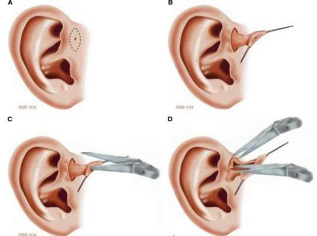
Figure 4. Inside-out technique. A: Making an elliptical incision around the fistula opening. B: Make stitches around hole fistula as handle For facilitate dissection channel fistula. C: Open channel fistula with scissors Which sharp. D: Using a magnifying glass, the methylene blue-stained wall (“inner”) and the outer wall of the fistula tract are freed from the surrounding tissue. 5
Another technique is a wide radical local excision defined as the supraauricular approach. In this technique, the elliptical incision around the sinus orifice is extended higher up into the pre- and supraauricular temporal areas. (Figure 6) This allows for better surgical visibility without adverse aesthetic consequences. 5

Picture 5. Approach supra-auricular And stitches bone helical cartilage . 5
Procedure closing classified become 3 category that is procedure involving primary closure alone, procedures involving primary closure with extensive undermining, and procedures involving auricular flap repair. Primary closure with extensive undermining is performed in the resection area. Which wide. Tension can avoided with weakening Which wide like a facelift procedure. Silicone drains are then inserted into the posterior side. The subcutaneous tissue is closed with primary sutures. 5
If bone vulnerable auricular open during excision And damage The network is extensive, closure is performed with posterior auricular transposition or rotation flap. The flap is moved into the defect with up to 120° rotation on the subcutaneous pedicle. (Figure 7). 5

Picture 6. Framework technique closing defect after excision preauricular sinus . 5
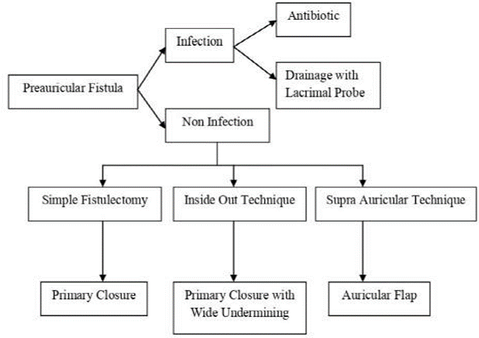
Picture 8. Algorithm reconstruction excision preauricular sinus . 5
In the study of Xu et al., 78 children with preauricular fistula infection were selected. In all children, a small spindle incision was made along the fistula, and then skin And network subcutaneous incised For show perichondrium of the helix of the ear and divided along the perichondrium to reveal the fistula and cyst. Almost all secretory and infected preauricular fistulas are attached on perichondrium helix ear. In case This, part bone The cartilage should be removed, and damage to the helical angle cartilage should be avoided as much as possible. In study This, time act carry on the longest is 3 year, And No recurrences were found. One child had a dead space due to sutures; hematoma formation and secondary infection occurred after drainage on the second postoperative day, but the child recovered after the area was cleaned. And sewn as well as installed bandage compression. Matter This Also showed that the surgical cavity was larger after the fistula tissue and granulation tissue were cut, especially in secretory preauricular fistula with infection. It is difficult to eliminate the dead cavity by suturing layer by layer, but intermittent sutures should be used. The middle part was sutured vertically to prevent varus. After surgery, the external auditory canal, concha cavity, scaphoid fossa, and triangular fossa were filled with gauze, and then a compression bandage was applied. If There is defect after network granulation issued from wound or used wound in front of the fistula, the subcutaneous tissue is separated and sutured with tension which is reduced, and a small drainage strip is installed in position. 7
Wide And Depth of Resection
In general anatomical, If A fistula experience obstruction, Can develops into a cyst due to accumulation of local secretions. The upper limit of this cyst is at the cranial junction of the ear, the lower limit is at the upper edge of the parotid gland, the anterior limit is at the growth margin, and the posterior limit is at the inner surface of the perichondrium. posterior helix ear. In general general, fistula And network granulation does not pass through the superficial layer of the temporal muscle fascia. This part has a safe border with the parotid gland and facial nerve, and is separated by the perichondrium helix ear during process resection For to expose bone helical cartilage. During the resection procedure, the lesion and granulation tissue are dissected along the superficial layer of the temporal muscle fascia downward toward the helical angle to cut the helical spine and the cartilage portion, as well as to posterior direction towards surface in perichondrium posterior helix ear. On stage infection, important For lift fistula And network inflammation together so that healing process can happen with optimal in implementation treatment first. 7
Repair Local
When fistula preauricular experience swelling repetitive, Good in purulent or local form, this indicates the secretory nature of the fistula. If lesi the contain network granulation inflammation chronic or object foreign body firmly embedded in the cartilage, patient healing can be difficult even with simple anti-infection treatment protocols and local dressing changes. In addition, the psychological impact on children and parents is also difficult to assess accurately. According to the report of Guo et al., plastic and cosmetic surgical interventions play a crucial role in the resection and repair of complex preauricular fistulas. During surgery, the incision design is adjusted according to the relationship between the focus of infection and the location of the fistula. Skin closure is also important to repair the wound, thus achieving optimal surgical results. Although fistula preauricular generally No need maintenance, Untreated infections can lead to recurrent infections and result in significant scarring after surgery. 7
The study by Scheinfeld et al., also emphasized the importance of surgical resection in cases of recurrently infected preauricular sinus. Postoperative management becomes crucial, as shown by the study by Tian et al., who recommended consideration of open drainage in case of abscess, and delivery specimen For culture bacteria. Study by Xu et et al., highlighted the importance of designing a small incision along the fistula to ensure a good cosmetic result. Preservation of the blood supply to the skin flap is also emphasized by sharp separation during the procedure. During flap formation, the subcutaneous tissue is carefully removed while the dermis of the flap is preserved as much as possible. Sewing skin done without tension For minimize risk of postoperative complications. 7
Common complications that often occur in cases of preauricular fistula and its surgical procedures include postoperative hematoma, wound dehiscence, wound infection, abscess, and facial nerve injury which is a more serious complication. 2

Picture 7. Photo pre-operative show hole fistula seen in area crus helix. 9
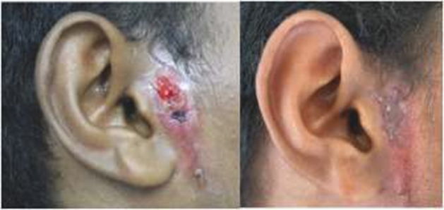
Figure 8. Preoperative photos (a) acute infection; (b) post-antibiotic administration (immediately before operation) appear hole preauricular in area preauricular. 9

Picture 9. Intraoperative photo . 9

Picture 10. Photo 2 month post operation. 9
SUMMARY
Preauricular fistula is a congenital abnormality of the soft tissues around the front of the ear. This condition can occur singly or multiple (unilateral or bilateral), with the possibility of multiple fistulas forming in one ear, especially in the auricle. Preauricular fistula is caused by incomplete fusion of the six auditory processes during preauricular development. Inadequate treatment can lead to recurrence, therefore, appropriate management often involves surgery, which in most cases can resolve the problem successfully.
REFERENCE
Vedas LPKE, Susanti IGAD. Abscess Sine Preauricular Here. Ganesha Med
J. 2023;3(1).
Tian CH, Chen XJ. Severe bleeding after surgery of preauricular fistula: A case report. World J Clin Cases. 2021;9(29):8932–7.
Li K, Hao Y, Zhao J, Zhou L, Wu Y, Zeng X, et al. Surgical treatment of preauricular fistulas: a 12-year single-center clinical observation. BMC Surg. 2023;23(1):1–8.
Park JY, Lee S, Kim HJ, Jung SG. Ectopic Preauricular Sinus in a Facial Cleft and Microtia Patient. Arch Craniofacial Surg. 2017;18(4):287–91.
Widodo DW, Harba'i HM. Surgical management of preauricular based on plastic-reconstructive algorithm. Auto Rhino Laryngol Indonesia. 2019;49(1):85.
Nagalingeswaran A, Kumar RD. Infra-auricular Sinus: A Very Rare Case Presentation. Indian J Otolaryngol Head Neck Surg. 2019;71(s2):1502–4.
Xu Y, Seng D, Jiang L, Wang S, Ni X, Zhang J, et al. Summary of the Experience in the Diagnosis and Treatment of Complex Preauricular Fistula in 78 Children. Front Surg. 2021;7(February):1–5.
Park H, Seong J, Park H, Yeo H. Standardized surgical strategy for the treatment of preauricular sinus to reduce recurrence. Arch Craniofacial Surg. 2023;24(5):223–9.
Widodo DW, Anatriera RA, The Goddess AP, Jonah RE. Recurrent preauricular fistula surgery guided by fistulography. J Pediatr Surg Case Reports. 2022;81.
Knipe H, Guardian T, Bell D. Preauricular sine. Radiopaedia. 2024.
Kumar Chowdary KVS, Sateesh Chandra N, Karthik Madesh R. Preauricular Sinus: A Novel Approach. Indian J Otolaryngol Head Neck Surg. 2013;65(3):234–6.
Hwang J, Cho J, Burm JS. Accessory auricle: Classification according to location, protrusion pattern and body shape. Arch Plast Surg. 2018;45(5):411– 7.
Matev B, Lyutfi E, Stoyanov GS, Sapundzhiev NR. Preauricular Sinus: A Tale of Forgetful Rediscovery. Cureus. 2020;12(6):6–13.
Vegetables CCAFM, Dickers FG, with baker BS. The Developmental Origin of the Auricula Revisited. Laryngoscope. 2020;130(10):2467–74.
Das C, Khaowas A. Pre-auricular Sinus with Post-auricular Extension: An Uncommon Variants. Indian J Otolaryngol Head Neck Surg. 2019;71(s2):1511– 4.
Comments