LITERATUR REVIEW
I Made Nudi Arthana
Disturbance Hearing
There is three type disturbance hearing, that is :
1 Conductive
Disturbance hearing conductive happen If wave voice No in a way adequately delivered through the outer and middle ear to vibrate the fluid in the inner ear. Possible causes are physical blockage of the ear canal by cerumen, rupture of the eardrum, middle ear infection with fluid accumulation or restriction of ossicular movement due to the bony attachment between the stapes and the oval window. 10
2 Sensorineural
In sensorineural hearing loss, sound waves are transmitted to the inner ear, but are not translated into nerve signals that can be interpreted by the brain as a sensation of sound. There are three recognizable patterns of sensorineural deafness, namely bilateral progressive loss , unilateral progressive sensorineural loss and sudden sensorineural deafness . 11
In bilateral progressive loss, cochlear degradation occurs due to aging, for example presbycusis, it can also be caused by ototoxic drugs or long-term exposure to excessive noise. Examples of ototoxic drugs include aminoglycoside antibiotics. Elderly patients and those with impaired kidney function are more susceptible to bilateral progressive loss. Long-term exposure to excessive noise can damage hair cells in the organ of Corti, usually occurring in industrial workers. shooter, usage tool electronic. Degrees severity depends on sound intensity, duration of sound exposure, individual resistance. 12
Unilateral progressive sensorineural loss always refers to Meniére's disease (endolymphatic hydrops), or acoustic neuroma. 15 Sudden sensorineural deafness is more common unilaterally, and may be caused by head trauma. or ear, infection viral (mumps, measles, varicella zoster) or sudden disturbance of cochlear circulation. Sudden sensorineural deafness can also refer to acoustic neuroma or barotrauma. 12
3 Mixture
This type of hearing loss is a combination of conductive hearing loss and sensorineural hearing loss. Initially, this type of hearing loss is a conductive type (eg otosclerosis), then develops further into a sensorineural disorder. It can also be the other way around, initially a sensorineural hearing loss, then accompanied by a conductive disorder (eg presbycusis), then infected with otitis media. Both disorders can occur together. For example, severe head trauma that simultaneously affects the middle ear and inner ear. 13
ISO ( International Standard Organization ) classifies deafness into several degrees (based on the hearing threshold in audiometric examinations), namely: 13
Normal, If threshold hearing on inspection audiometry range between 0-25 dB
Deaf light, If threshold hearing on inspection audiometry ranges between >25-40 dB
Moderate deafness, if hearing threshold in audiometric examination ranges between >40-55 dB
Deaf currently heavy, If threshold hearing on inspection audiometry ranges from >55-70 dB
Deaf heavy, If threshold hearing on inspection audiometry ranges between >70-90 dB
Deaf very heavy, If threshold hearing on inspection audiometry ranges between >90 dB
4 Presbycusis
2 Definition and Epidemiology
Presbycusis originate from Language Greece that is prebys It means age, And ákousis is hearing. Presbycusis is a hearing loss that accompanies the aging process. Pure tone audiometry shows a picture of bilateral symmetrical hearing loss that begins at high tones and is sensorineural in nature with no underlying abnormalities other than the general aging process. 14
According to the World Health Organization (WHO), currently an estimated 360 million (5.3%) people in the world suffer from hearing impairment, 328 million (91%) of whom are adults (183 million men, 145 million women) and 32 million (9%) are children. The prevalence of hearing impairment increases with age. 14
In the United States, hearing loss is common in nearly two-thirds of adults aged 70 years and older, with the highest frequency in men and degrees disturbance hearing most is degrees light. On research in Iran Also show sufferer presbycusis most is group men over 60 years old. The most common type of presbycusis is the sensory type, followed by the neural, conduction and metabolic types. Likewise, research conducted at Adam Hospital Malik Medan get results Which The same in the form of patient presbycusis The most people who came were in the group above or equal to 70 years of age with the highest frequency in the male group. 14,15 Sogebi also conducted the same research in Nigeria and obtained results in the form of male sufferers more Lots from Woman with group age most 71-80 year. The highest degree of hearing loss is moderate and the highest type of presbycusis is strial. 15
In a study on the characteristics of presbycusis sufferers at Dr. Hasan Sadikin General Hospital, Bandung, there were sufferers with a group of boys higher than Woman. Type presbycusis most is type neural, followed type sensory, type metabolic/strial And type mechanical/conduction cochlear. Looks number The incidence of presbycusis is most common in people aged > 65 years. The degree of hearing loss in most presbycusis sufferers is mild. 16
Study Which The same Also done in General Hospital Sanglah Denpasar Which obtained results in the form of presbycusis sufferers most often found in men with the largest age range being 60-70 years, and the type of presbycusis most often found is the strial type. 17
5 Pathology
Process degeneration cause change structure cochlea And nerve VIII. In the cochlea, the most striking changes are atrophy and degeneration of the supporting hair cells in the organ of Corti. The atrophy process accompanied by vascular changes also occurs in the stria vascularis. In addition, there are also changes in the form of a reduction in the number and size of ganglion cells and nerves. The same thing also happens to the myelin of nerve axons. 17
6 Factor Risk
Presbycusis allegedly relate with a number of factor risk, between other :
Age And gender
Presbycusis occurs on average at the age of 60-65 years and above. The effect of age on disturbance hearing different between man And Woman. Men experience more hearing loss at high frequencies and only a slight decrease at low frequencies when compared to women. The difference type sex on threshold hear frequency tall This because men are generally more exposed to noise in the workplace than women. Sunghee et al. stated that the difference in the influence of gender on presbycusis is not entirely due to changes in the cochlea. Women have smaller earlobes and ear canals which can cause a masking effect on low-frequency noise. Pearson stated that hearing sensitivity is better in women than in men. 18
Hypertension
Long-term hypertension can increase vascular resistance, resulting in dysfunction of blood vessel endothelial cells accompanied by increased viscosity. blood, decline flow blood capillary And transport oxygen. Matter This causes damage to auditory cells so that the signal transmission process is disrupted, causing communication disorders. Sensory neural hearing loss can occur due to microcircular insufficiency of blood vessels such as embolism, bleeding, or vasospasm. 18
Diabetes mellitus
On patient with diabetes mellitus (DM), glucose Which bound on Proteins in the glycosylation process will form advanced glycosylation end products (AGEP) which accumulate in the tissue and reduce the elasticity of the blood vessel walls (arteriosclerosis). The next process is that the blood vessel walls become thicker and the lumen narrows, which is called microangiopathy. Microangiopathy on the organ cochlea will cause atrophy and decrease hair cells, when condition This happen on vase nerve VIII, ligament And ganglion spiral in Schwann cells, myelin degeneration, and axon damage will cause neuropathy. The National Health Survey USA reported that 21% of diabetics suffer from presbycusis, especially at the age of 60-69 years. The audiometry results of DM patients showed that the frequency of hearing loss in this group was higher when compared to patients without DM. 18
Smoking habit
Cigarettes contain nicotine and carbon monoxide which have the effect of disrupting blood circulation, are directly ototoxic, and damage the nerve cells of the cochlea. Carbon monoxide causes ischemia through the production of carboxy-hemoglobin (a bond between CO and hemoglobin) so that hemoglobin becomes No efficient tie oxygen. Bond between hemoglobin with CO much stronger than hundreds of times compared to oxygen. As a result, there is a disruption in the supply oxygen to the organ of corti in the cochlea and causes ischemic effects. In addition, other effects of carbon monoxide are spasms blood vessels, blood viscosity, and arteriosclerotic. Insufficiency of the cochlear blood circulation system caused by smoke become reason disturbance hearing on frequency tall progressive. The blood vessels that supply blood to the cochlea do not have collaterals so they do not provide an alternative blood supply through other routes. 19
In a study conducted by Dawes et al (2014), active smokers and passive smokers were associated with increased hearing loss. Cruichksanks' study reported that non-smokers who lived with smokers more at risk experience disturbance hearing compared to they who live with non-smoking family members. 20
Mizoue et al. studied the effects of smoking and noise on hearing loss through health examination data of 624 steel factory workers in Japan. The results showed a significant picture of hearing loss at high frequencies due to smoking with a risk three times greater . 20
History noisy
Noise-induced hearing loss is a sensorineural hearing loss that is initially not noticed because it does not interfere with daily conversation. Risk factors that influence the severity of deafness are the intensity of the noise. noisy, frequency, long exposure per day, long time Work with noise exposure, individual sensitivity, age, and other factors that may influence. Based on this, it can be understood that the amount of noise energy exposure received will be proportional to the damage obtained. This is because continuous exposure can damage the cochlear hair cells. 19
7 Classification
Based on audiometric descriptions and pathological changes that occur, in 1969 Schuknecht et al classified presbycusis into 4 categories, namely sensory, neural, metabolic (strial presbycusis) and mechanical (cochlear presbycusis).
Sensory
Presbycusis sensory originate from degeneration organ Corti Which started from basal and progresses gradually towards the apex. High frequency hearing is impaired but speech discrimination remains good. Sensory presbycusis is also caused by damaged outer hair cells. According to the Schuknecht Classification, the occurrence of presbycusis sensory donate 5% from total case presbycusis. The incidence of sensory presbycusis was also not high in the study by Gates et al. 21
Neural
Neural presbycusis refers to the loss of neuronal cells in the cochlea. Otte, et al. showed that approximately 2100 neurons are lost every 10 years in humans. Loss of 50% of the afferent nerves results in reduced speech discrimination, and 90% loss results in changes in hearing thresholds. 22
Metabolic
Presbycusis metabolic or strial presbycusis due to by atrophy stripe vascularis, loss 30% or more network in stripe vascular cause decline
hearing threshold. Mills said metabolic type is the main cause presbycusis. History family influential. On audiogram looks looks flat but speech discrimination is still good. 21,22
Mechanics
Mechanical presbycusis occurs due to degenerative changes that cause stiffness. in area membrane basilaris so that hinder its movement. The audiogram shows sloping and no disturbance in speech discrimination. 22
8 Symptom Clinic
The main complaint of presbycusis is a gradual and progressive hearing loss, symmetrical in both ears. When the hearing loss occurs is not known for sure. 23 Another complaint is ringing in the ears (high-pitched tinnitus). Patients can hear the sound of conversation, but have difficulty understanding it, especially when spoken quickly in a place with a noisy background ( cocktail party deafness ). If the sound intensity is increased, pain will arise in the ear, this is caused by the factor of nerve fatigue (recruitment). Usually patient Which come, sigh difficulty in understand the conversation rather than complaining about not being able to hear. 24
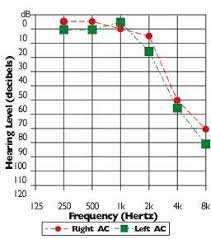
Picture 1. Pattern Tone Audiometry Pure on Presbycusis 24
9 Diagnosis
The diagnosis can be made by a detailed history, physical examination, and hearing tests including an audiogram. The history of hearing loss is often difficult. A common clinical complaint is “my hearing is fine. It’s just that my wife/husband forces me to come in for a hearing test.” Patients also often have difficulty explaining when they first noticed their hearing loss. This event is usually associated with when they were first unable to communicate over the telephone.
Patient Also often complain about existence tinnitus. Tinnitus defined as an abnormal ringing sound in the ear. Tinnitus patients with sensorineural etiology will describe the tinnitus as being worse in a quiet environment where there are no other disturbing sounds. Patients often complain that tinnitus very bother on moment approaching Sleep or wake up. 13 With inspection otoscopic, looks membrane timpani gloomy,
its mobility reduce. On test tuner obtained deaf sensorineural. Pure tone audiometry examination showed a high tone, bilateral and symmetrical nerve deafness. 13
Earwax should be cleaned before ear inspection. Earwax can block the ear canal and cause hearing loss in the elderly and reduce the quality of hearing aid use. Presbycusis is usually diagnosed when the patient has the following criteria: symmetrical increase in hearing threshold, no trauma, use of ototoxic drugs, history of ear disease and ear surgery, and minimal conductive hearing loss (10 dB or lower) and age over 65 years. 11
10 Treatment And Prevention
Presbycusis cannot be cured. Rehabilitation as an effort to restore hearing function is carried out by installing a hearing aid. 30 Sometimes the installation of a hearing aid needs to be combined with speech reading training and auditory training. the training procedure is carried out together speech therapist. 25 For prevention against hearing loss can be done with sufficient vitamin intake. Vitamin intake is important for normal hearing function. A survey of the general population in the United States Union aged 20-69 year find that diet tall vitamin C, beta carotene, and magnesium are correlated with better hearing. 26
Although presbycusis difficult prevented Because is change degenerative, prevention of aggravating factors can be an important factor in reducing incident presbycusis. For example, with avoid use ototoxic drugs (antibiotics such as aminoglycosides or chemotherapy drugs). Stop or reduce smoke And avoid exposure cigarette passively Also helps prevent further hearing loss. 27
REFERENCE
Wang J, Puel JL. Presbycusis: An Update on Cochlear Mechanisms and Therapies. J Clin Med. 2020 Jan;9(1):218.
Paplou V, Schubert NMA, Pyott SJ. Age-Related Changes in the Cochlea and Vestibule: Shared Patterns and Processes. Front Neurosci. 2021 Sep 3;15:680856
Kim TS, Chung JW. Evaluation of Age-Related Hearing Loss. Korean J Audiol 2013; 17 : 50-53.
Lee KY. Pathophysiology of Age-Related Hearing Loss (Peripheral and Central). Korean J Audiol 2013; 17: 45-49.
Hall, J. 2016. Guyton and Hall Textbook of Medical Physiology, 13th edn, Elsevier: Philadelphia.
Hansen, J. 2014. Netter's Clinical Anatomy, 3rd edn. Elsevier: Philadelphia.
Howarth, A. 2005. Ageing and the auditory system. Postgraduate Medical Journal. 82(965), hal. 166–171.
Ito, J. 2015. Regenerative Medicine in Otolaryngology. Springer: Japan.
Ministry of Health of the Republic of Indonesia. 2013. Healthy Hearing for a Happy Life.
Khan, B. H., Hello, S. and Palous, P. (2012). Pattern of Pure Tone Audiograms in Presbyacusis. pp. 84–87.
Kim, T. S. & Chung, J. W. 2013. Evaluation of age-related hearing loss. Korean Journal of Audiology. 17(2), hal. 50–53.
Lee, K.-Y. 2013. Pathophysiology of Age-Related Hearing Loss (Peripheral and Central). Korean Journal of Audiology. 17(2), hal. 45.
Levine, S. 1997. Inner Ear Disease. BOEIS: Book Teaching ENT Diseases, 6th Ed. EGC: Jakarta.
Lin, F., Thorpe R., Gordon-Salant,S. et al. 2011. Hearing Loss Prevalence and Risk Factors Among Older Adults in the United States. The Journals of Gerontology Series A: Biological Sciences and Medical Sciences. 66A(5), hal. 582–590.
Liu, X. & Yan, D. 2007. Ageing and hearing loss. The Journal of pathology. 211, hal. 188–197.
Ludman, H. 2007. ABC of Ear, Nose and Throat, 5th edn. Blackwell: Australia.
Munir, N., & Clarke, R. 2013. Ear Nose and Throat at a Glance. Wiley Blackwell: West Sussex.
Muyassaroh. 2012. Faktor risiko presbikusis. Journal Medical Assocation. 62(April), hal. 155– 158.
Nagel, P., & Gurkov, P. 2012. Basics of ENT Science, 2nd Edition. EGC: Jakarta.
Nuryadi, NKR, Wiranadha, M. and Sucipta, W. 2017. Characteristics of presbycusis patients in the Polyclinic ENT-KL General Hospital Sanglah Denpasar 2013-2014. Medicina. 48(1), p. 46.
Rolim, LP et al. 2017. Effects of diabetes mellitus and systemic arterial hypertension on elderly patients hearing. Brazilian Journal of Otorhinolaryngology.
Sarafraz, M., Saki, N., Maleki, M., et al. 2015. Distribution of audiometric findings in patients with presbycusis. Biomedical and Pharmacology Journal. 8(SEMAR), p. 37-41.
Sherwood, L. 2011. Human Physiology: from Cells to Systems, 6th Edition. EGC: Jakarta.
Soetirto, I., Hendarmin, H., & Bashiruddin, J. 2012. Hearing Disorders and Ear Disorders. Book Teaching Ear, Nose, Throat, Head and Neck Health Sciences, 7th Edition. FKUI Publishing Agency: Jakarta.
Suwento, R., & Hendarmin, H. 2012. Hearing Disorders in Geriatrics. Book Teach Knowledge Health Ear Nose Throat Head And Neck, 7th Edition. FKUI Publishing Agency: Jakarta.
Tortora, G., & Derrickson, B. 2012. Principles of Anatomy and Phsiology, 13th edn. Quad Graphics: United States.
Yang, C.-H., Schrepfer, T. dan Schacht, J. 2015. Age-related hearing impairment and the triad of acquired hearing loss. Frontiers in Cellular Neuroscience, 9(July), hal. 1–12.
Ario M M, Anggraeni R, Aroeman N A. 2022. Karakteristik Penderita Presbikusis di Kota Bandung Tahun 2019. Journal of Medicine and Health Vol. 4 No. 1 February 2022. e-ISSN: 2442-5257
Kim SH, Lim EJ, Kim HS, Park JH, Jarng SS, Lee SH. Sex differences in a cross sectional study of age-related hearing loss in korean. Clin Exp Otorhinolaryngol. 2010;3(1):27–31.
Yamasoba T, Lin FR, Someya S, Kashio A. Current concepts in age-related hearing loss: epidemiology and mechanistic pathways. NIH Public Access. 2013;23(1):1–7.
Mondelli GC, Lopes CA. Relationship between Arterial Hypertension and Hearing Loss. Intl.Arch. Otorhinolaryngol. 2009;13:63-8.
Nuryadi, NKR, Wiranadha, M. and Sucipta, W. 2017. Characteristics of patients with presbycusis in the Polyclinic THT-KL RSUP Sanglah Denpasar year 2013-2014. Medicine. 48(1), p. 46.
Gates GA, Mills JH. Presbycusis. The Lancet. 2005;366(9491):1111-20.p.1-10
Comments