PSEUDOANGINA LUDOVICI
I Made Nudi Arthana
LITERATUR REVIEW
Anatomy Neck
Pseudoangina Ludovici is an abscess that forms in potential space between the deep neck fascia as a result of the spread of infection from various sources such as teeth, mouth, throat, paranasal sinuses, ears and neck. In the neck area, there are several potential spaces that are limited by fascia cervical. Fascia cervical shared become two that is fascia cervical superficial and deep. Fascia cervical superficial located in lower dermis And consists of from fibroadipose tissue. This fascia envelops sensory nerves, superficial blood vessels, lymph nodes, platysma muscle and mimic muscles. 4
The deep cervical fascia consists of fibrous connective tissue and is divided into three layers, namely the superficial, medial and deep layers. The superficial layer of the deep fascia is also called the investing layer. Rule of two of the superficial layer This is wrap two the muscles that located on bone hyoid, namely the masseter muscle and the anterior venter of the digastric muscle; two neck muscles, namely the trapezius And muscular sternocleidomastoideus; two gland saliva namely the parotid and submandibular glands; two spaces namely the parotid and masticator spaces. 4
Layer media fascia deep Which called Also fascia cervical consists of from the muscular and viscera divisions. The muscular division encloses the muscular sternohyoid, muscle sternothyroid, muscular thyrohyoid And muscular omohyoid. Division The viscera surround the parathyroid glands, thyroid gland, esophagus, larynx, pharyngeal constrictor muscles and buccinator muscles. 4
The deep layer of the deep fascia is also called the prevertebral fascia, and consists of the alar and prevertebral divisions. The alar division is located between the buccopharyngeal fascia anteriorly and division prevertebra in posterior. Division alar is the anterior wall of the danger space . This fascia extends from the base of the skull to the thoracic vertebrae. second. Division prevertebra is located in front vertebral column and extends to the lateral pass prevertebral muscles and Then fuse with transverse processes and their accompanying ligaments. This fascia forms the anterior wall of the prevertebral space and the posterior wall of the danger space . 4
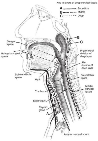
Picture 1. Piece midsagittal neck 4
The potential space of the deep neck is divided into spaces that encompass the entire neck length, space suprahyoid And infrahyoid. The space involved along the neck consists from retropharyngeal space, danger space , prevertebral space and carotid space. The retropharyngeal space extends from the base of the skull to the tracheal bifurcation in the mediastinum. superior. Space retropharyngeal bordering with sheath carotid on the lateral side, buccopharyngeal fascia anteriorly and the alar division of the prevertebral fascia posteriorly. Danger bordered space with retropharyngeal space in anterior and prevertebral space posteriorly, extending from the base of the skull to the diaphragm. The prevertebral space extends from the base of the skull to the coccygeus, bordering the danger space anteriorly, the vertebral bones posteriorly and the transverse processes laterally. The carotid space or also called the visceral vascular space is a potential space in in sheath carotid. In in this space there is artery carotid, internal jugular vein, vagus nerve and sympathetic plexus. 4
The suprahyoid space consists of the submandibular, parapharyngeal, parotid, masticator, and peritonsil And temporal. Submandibular space located between the mucosa of the floor of the mouth and the superficial deep fascia at the bottom. This space is bounded by the hyoid bone posteroinferiorly, the mandible anteriorly and laterally and the base of the tongue posteriorly. This space is divided incompletely by the mylohyoid muscle become room sublingual on part on And submandibular space And submental at the bottom. The submandibular and submental areas are separated by the anterior venter of the digastric muscle but these two areas communicate freely with each other. The submental space is bounded by the hyoid bone inferiorly, the mandible superiorly and the anterior venter of the digastric muscle on both sides lateral. Submental space containing jugular vein anterior, submental lymph nodes, mylohyoid muscle and nerve, submental branches of the facial artery and facial vein. 4
In the parotid space there are the parotid glands, lymph vessels, external carotid artery, superficial temporal artery, posterior facial vein, facial nerve. And nerve auriculotemporalis. Layer superficial fascia deep separates around the mandible to form the masticator space. In this space are the masseter muscle, medial and lateral pterygoid muscles, ramus and corpus of the mandible, and temporalis tendon. The masticator space consists of the masseter space and the pterygoid space. The masseter space is located between the ramus of the mandible and the masseter muscle while the pterygoid space is located between the ramus of the mandible and the pterygoid muscle. This space is located anterior and lateral to the parapharyngeal space and inferior to the temporal space. 4
Room peritonsil located lateral from capsule tonsil And medial from constrictor muscle superior. Arch palatoglossus And palatopharynx to form limit anterior and posterior room This. On part inferior restricted by 1/3 part posterior tongue. The temporal space is located between the temporal fascia on the lateral side and the periosteum part squamous OS temporal on part medial. Muscle temporalis separates this space into superficial and deep spaces. The infrahyoid space consists of the anterior visceral and suprasternal spaces. The anterior visceral space or called Also room pretracheal located on neck front from cartilage thyroid downwards to the superior mediastinum at the level of the fourth thoracic vertebra. The suprasternal space is located in the superior sternal notch , covered by the superficial layer of deep fascia. 4
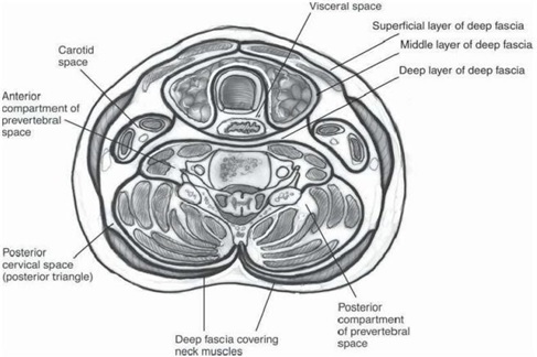
Picture 2. Cuts transverse neck as high as Thyroid 4
Definition
Pseudoangina ludovici can be a continuation of angina lodovici, where there is fluctuation in palpation. Angina ludovici is a submandibular space infection in the form of rapidly spreading cellulitis, potentially causing death, affecting the sublingual and submandibular spaces. Generally, the infection begins with cellulitis, then develops into fasciitis and finally develops into an abscess that causes suprahyoid induration, swelling on base mouth And elevation as well as change location tongue posteriorly. 5
Epidemiology
Pseudoangina ludovici is a rare disease that can be potentially life-threatening if the infection spreads to the mediastinum. The main predisposing factor is poor orodental hygiene while the predisposing factor other is systemic disease And immunodeficiency disease because these diseases can facilitate the development of bacteria and the spread of infection, such as diabetes mellitus, neutropenia, alcoholism, aplastic anemia, glomerulonephritis, dermatomyositis and systemic lupus erythematosus. incident pseudoangina ludovici most often on age 20 until 60 year, with male dominance compared to females being 3:1. The death rate due to pseudoangina ludovici before known to him antibiotic reach number 50% of all reported cases, in line with the development of antibiotics, treatment surgery Which Good And action Which fast And appropriate, so At the moment the mortality rate is only 5%. 6
Etiopathogenesis
The most common cause of this abscess is a tooth infection. Pulp necrosis due to untreated deep caries and deep periodontal pockets are the pathways for bacteria to reach the periapical tissue. Because of the large number of bacteria, the infection that occurs will spread to the spongiosa to the cortical bone. If this bone thin, then infection will penetrate and enter the soft tissue. Distribution infection This depends from Power stand network body. Odontogens can spread through connective tissue, blood vessels blood And lymph vessels. The most common is percontinuitatum because there is a gap or space between the tissues that has the potential to be a place for pus to collect. 7,8 The course of infection in the upper jaw can form palatal abscess, submucosal abscess, gingival abscess, cavernous sinus thrombosis, labial abscess and facial abscess. The course of infection in the lower jaw can form sublingual, submental, submandibular abscess, submaseteric abscess and pseudoangina ludovici. root molar second And third located in behind lower line mylohyoid which is located in aspect in mandibular, so that If molar second or third infected and forms an abscess, the pus will spread to the submandibular space and can extend to the parapharyngeal space. 7
Pseudoangina ludovici caused by odontogenic infection, originates from lower second or third molars. This tooth has the root above the mylohyoid and abscesses in this location can spread to the submandibular space. Infections that spread beyond the root of the tooth originating from the premolar tooth are generally located in the first sublingual while infections outside the root of the tooth originating from the molar tooth are generally located in the submandibular space. 8 The infection rapidly spreads from the submandibular, sublingual and submental spaces. Which cause swelling And elevation tongue from base mouth. The potential space for pseudoangina Ludovici to occur is the suprahyoid space which is located between the muscles that attach the tongue to the hyoid and mylohyoid bones. Inflammation in this space causes swelling of the tissues of the floor of the mouth. And push tongue to on And behind, so that cause airway obstruction. 8
Infection in the submental space is usually limited by the rigid union of the deep cervical fascia with the anterior digastric muscle and the hyoid bone. in the submaxillary space is usually limited in in the space itself, but can also follow along the submaxillary duct of Wartoni and follow the glandular structure towards the sublingual space or can also extend downwards along the hyoglossus muscle towards the fascial spaces of the neck. 9
In sublingual space infections, edema is present in the weakest area of the sublingual space. superior and posterior, so as to encourage supraglottic larynx And tongue backwards, eventually narrowing the channel and obstructing the airway. The spread of infection ends in the anterior part, namely the mandible and in the inferior part that is m.mylohyoid. Process infection Then walk in part superior and posteriorly, extending to the floor of the mouth and tongue. The hyoid bone limits this process inferiorly so that the swelling spreads to the front of the neck causing changes in shape and a “bull neck” appearance. 9,10
Manifestation clinical
Extra oral clinical symptoms include erythema, swelling, hardness to the touch. like board, elevation temperature neck And dysphonia or hot potato voice due to edema in the vocal organs. Intra-oral clinical symptoms include swelling, pain and elevation tongue. Other symptoms include presence of swelling the painful one on the floor of the mouth and anterior part of the neck, fever, pain on swallowing or odynophagia, hypersalivation, trismus, pain in the teeth, hoarseness, stridor, respiratory distress and cyanosis. 9 On inspection physique there is fever with characteristics base the mouth that swollen and reddish. Found caries on tooth molar lower, induration and swelling of the submandibular space which may be accompanied by the tongue being pushed forward on. Trismus can occur Because existence irritation on m. masticator. Signs important like patient unable to swallow his saliva Alone, dyspnea, tachypnea, stridor inspiration And cyanosis Which show existence blockage road breath. In patients, dysphonia may occur due to edema of the vocal structures. 10
Diagnosis
The diagnosis of pseudoangina ludovici is based on anamnesis, physical examination and supporting examinations.
Anamnesis
On anamnesis, obtained symptom beginning in the form of painful on area infected teeth, the chin feels tense and painful when moving the tongue. The patient may experience difficulty opening the mouth, speaking and swallowing, resulting in constant drooling and difficulty breathing. 11
Inspection Physique
On physical examination, the floor of the mouth is reddish and swollen. When the infection spreads to the back of the mouth, inflammation of the floor of the mouth will cause the tongue to be pushed up and back, blocking the airway. If the larynx is also swollen, a stridor sound will be heard when breathing. Swelling in the anterior neck tissue above the hyoid bone is often called a bull's neck appearance . Usually sufferer will experience dehydration consequence lack of fluid that is drunk and also food Which eaten. Fever tall Possible found, indicating the presence of systemic infection. There are 4 cardinal signs of pseudoangina ludovici, namely bilateral or more deep neck space involvement, gangrene accompanied by serosanguinous pus, involvement of connective tissue, fascia and muscles but not affecting glandular structures and distribution through facial space more often than through the lymphatic system. 11
Inspection Support
Supporting examinations that can be carried out include laboratory and radiology. On inspection blood edge obtained improvement leukocytes that identify existence infection I. Inspection culture And Sensitivity is carried out to determine the type of bacteria and the selection of antibiotics. Which in accordance. Photo plain network soft neck anteroposterior and lateral is procedure diagnostic Which important. On inspection soft tissue photos of the neck in both positions can provide an image of the deviation trachea, air in area subcutaneous, fluid in in network soft and swelling of the soft tissue area of the neck. The limitation of this examination is that it cannot differentiate between cellulitis and abscess formation. Chest X-ray is used to diagnose pulmonary edema, pneumothorax, pneumomediastinum or enlargement gland sap hilus clear . 11,12
A panoramic photo of the jaw can help determine the focal location of infection. or abscess, as well as structure bone jaw Which infected. Ultra examination sonography (USG) is means support diagnostic Which non-invasive and relatively inexpensive. Ultrasound can show the location and size of the pus as well as guide at the time of aspiration or abscess drainage. CT-Scan examination can help illustrate location And expansion abscess. On the CT-Scan image, low-density areas, increased contrast in the abscess wall and soft tissue edema around the abscess can be found. Magnetic resonance imaging (MRI) examination provides description improvement density on network Which experiencing inflammation compared to with network normal. Inspection MRI relatively expensive and not every hospital has this tool, so MRI examination in cases of pseudoangina ludovici is not a standard procedure. 11,12
TREATMENT
Assessment of the patient's general condition is important in the management of Ludovici's pseudoangina. The main priority is stabilization of the airway and circulation. Because pseudoangina ludovici has the potential to be life-threatening, patients should treated in House Sick. On case pseudoangina ludovici with road blockage breath required Handling quick with tracheostomy Which aiming to secure the airway. 13Antibiotic dose tall And spectrum wide given in a way intravenous for organisms grams positive And grams negative as well as germs aerobic And anaerobic. Antibiotics are given according to the culture and sensitivity results of the pus. Antibiotics used are high-dose penicillin-G and metronidazole, clindamycin, ceftriaxone and amoxicillin-clavulanic acid. Although still controversial, corticosteroids are given to reduce edema and increase antibiotic penetration. Intravenous anti-inflammatory drugs are given within 48 hours to reduce edema and protect the airway. 13
In pseudoangina ludovici, exploration is also performed for the purpose of decompression (reducing tension) and evacuation of pus or necrotic tissue. Surgery can be performed through an incision in the midline horizontally at the level of hyoid bone (3-4 fingers) under the mandible) aims to reduce the tension that formed on base mouth. Before done incision And drainage, Preparation for the possibility of tracheostomy should be done due to the inability to intubate the patient. Drainage incision is indicated if there is a suppurative infection with a radiological examination showing a picture of fluid accumulation in the soft tissue And crepitation. Drainage Also indicated If No There is repair after giving therapy antibiotics. Drainage was performed in the mylohyoid muscle into the sublingual space. Extracting the infected tooth is also important for the drainage process. 14
Antibiotics should be given immediately in adequate doses parenterally. Before there are culture and resistance results, antibiotics are given based on experience/in a empirical type germs Which often found that is germs aerobic and anaerobic. For the administration of drug therapy in patients with suspected pseudoangina ludovici can given antibiotics penicillin G 300,000-1,200,000 units/day or amoxicillin 25-30 mg/kgBW/day or cephalosporin 25-30 mg/kgBW/day or gentamicin 20-80 mg 1-2 times daily or clindamycin 600-900 mg intravenously every 8 hours or a combination of penicillin and metronidazole. Metronidazole can be given intravenously 3 x 500 mg/day. Antibiotic administration can reduce mortality from deep neck space infections, but infections in deeper spaces can cause fatal and life-threatening complications. 14If the abscess has spread to the deep neck space such as the parapharyngeal or retropharyngeal area, antibiotics need to be given according to culture and sensitivity tests and surgery. The decision to perform surgery is based on several considerations, namely if there is no improvement in 24 hour time which is marked with fever, pain pressure, persistent swelling and leukocytosis, threat of airway obstruction, presence of life-threatening neurovascular complications, pus appears more than 3 cm on CT-Scan. 15
Complications
The most common complications are airway obstruction, spread of infection And abscess to room neck in other And mediastinum as well as sepsis that causes the more difficult it is to handle and even can cause death. If there is spread to the intracranial area, it can cause cavernous sinus thrombus, meningitis and brain abscess. 7Other complications include asphyxia caused by edema in the soft tissue neck, infection wall carotid, rupture arteries, thrombophlebitis suppurative of the jugular veins, acute mediastinitis, emphysema, pleural effusion, mandibular osteomyelitis, bacterial pericarditis and aspiration pneumonia. 7
Prognosis
Prognosis of pseudoangina ludovici depends on the speed of airway protection And Then giving antibiotic. Pseudoangina ludovici can be fatal because it is life-threatening. Mortality in the pre-antibiotic era was around 50%. However, with early diagnosis, immediate airway protection, adequate intravenous antibiotics, and treatment in the intensive care unit (ICU), this disease can be cured without causing complications. Thus the mortality rate also decreases to less than 5%. 15
REFERENCE
Novialdi, Pulungan MR. Deep neck abscess germ pattern. Department of ENT-KL, Faculty of Medicine, Andalas University. 2010. Downloaded from url: http://repository.unand.ac.id/18384. Accessed on May 24, 2024.
Brotfain E, Koyfman L, Odes LS, Borer A, Refaely Y, Klein M. Case Report: Deep Neck Infection and Descending Mediastinitis as a Complication of Propionibacterim acnes Odontogenic Infection. Israel. 2015. Available at:http:
Rocha FS, Batista JD, Silva CJ, Júnior RB, Raposo LHA. Considerations for the Spread of Odontogenic Infections Diagnosis and Treatment 2015 2015-04- 22.
Faith M. Evaluation Management Abscess Neck In In Department ENT- KLHome Sick Hassan Sadikin Bandung Period January 2012– December 2012. Juke Unila. 2015; 5(9):33-37
Huang SH, Yang SW, Lee MH, See LC, Chen TM, Chen TA. Deep Neck Abscess: An Analysis of Microbial Etiology and The Effectiveness Of Antibiotics. Infection andDrug Resistance. 2008:1 :1
Forehead IM, Ersil P, Caminiti M. A Rare Complications of Teeth Abscess-Ludwig's Angina and Mediastinitis. In J Can Dent Assoc. America. 2001: p. 324-7
Higler Boies A. Oral Cavity and Pharynx. In: Textbook of ENT Diseases. Jakarta: EGC;1997. p.345-6.
Fachruddin D. Deep neck abscess. In: Iskandar M, Soepardi AE editor. Textbook of ear, nose, and throat diseases. 7th edition. Jakarta: Balai Penerbit FK-UI.2007: p. 185-8
Scott BA, Steinberg CM, Driscoll BP. Infection of the deep Space of the neck. In: Bailey BJ, Jhonson JT, Kohut RI et al editors. Otolaryngology Head and neck surgery. Philadelphia: JB. Lippincott Company. 2001.p.701-15 http://dx.doi.org/10.1155/2015/19013
Ballenger J J. Diseases of the oral cavity. In: Ballenger JJ, Snow Jr JB, eds. Otorhinolaryngology: Head and Neck Surgery. 15th Ed. United States of America :Williams & Walkins; 1996. p.233-234.
Novialdi, Triana W. Multiple deep neck abscesses with intubation difficulties and complications of pharyngocutaneous fistula. Department of ENT-KL, Faculty of Medicine , Andalas University . 2011. Downloaded from url: http://repository.unand.ac.id/18174. Accessed on May 24, 2024.
shutter K, Rathore PK, Wow V, Kumar S. Deep Neck Infections: Continuing Burdenin Developing World. International Journal of Phonosurgery and Laryngology.2013;3(1):6-9.
Rizzo P, Mosto MCD. Submandibular Space Infection: A Potentially Lethal Infection. International Journal of Infectious Diseases. 2009;13:327-33.
Oliver ER, Gillespie MB. Deep Neck Space Infections. In: Flint PW, Haughey BH, Lund VJ, Niparko JK, Richardson MA, Robbins KT, et al., editors. CummingsOtolaryngology Head and Neck Surgery. 5th ed. Philadelphia: Mosby, Inc.; 2010. p.201-8.
Aynehchi BB, Har El G. Deep neck Infection. In: Johnson JT & Rose CA, eds. Bailey's Head and Neck Surgery Otolaryngology, 5th edition, Philadelphia: Lippincort William & Wilkins, 2014: p. 749-816
Comments