Zygomatic Fracture
I Made Nudi Arthana
In zygomatic fractures, the location of the fracture depends on level its severity, Which involving four location, that is: rim orbit lateral and inferior, support ZMC, And arch zygomatic.
LITERATUR REVIEW
1 Epidemiology and Etiology Zygomatic Fracture
In zygomatic fractures, The zygomatic bone forms the lateral aspect of the midface and consists of the lateral and inferior orbital rims and the malar eminences, thus creating the width and projection of the face. 8,9 Fractures of the zygomatic arch occur in 10% to 15% of all fracture face And usually due to by blow direct to face. 10 Complex fracture zygomaticomaxillary (ZMC) occurs at around 25%-40% Of all facial fractures, the second most common after closed nasal fractures and are generally the result of traffic accidents, industrial accidents, sports injuries, and interpersonal violence. 11
The presence of aggravating factors such as bone conditions that experience osteoporosis also affect the incidence of maxillofacial fractures including zygoma fractures. This was obtained from a retrospective study conducted on 59 patients. fracture maxillofacial Which aged 60 year to on in A trauma center in Ohio United States for 11 years, from 1989 to 2000. The more severe the osteoporosis condition, the greater the possibility of the number of maxillofacial fractures experienced. Therefore, a lighter impact due to a fall can cause multiple maxillofacial fractures as occurs in motor vehicle accidents if the patient has severe osteoporosis. 12,13
In children the overall prevalence of facial bone fractures is much lower than in adults. Approximately 5-15% of all facial fractures occur in child. Prevalence fracture face pediatrics most low on baby And increase progressively according to increasing age. There are 2 age peaks where the frequency of fractures is highest in pediatrics, namely between the ages of 6-7 years related to the age of starting school and at the age of 12-14 years related to increased physical activity and participation in sports during puberty and adolescence. The mechanism of facial trauma including zygoma in children is similar to that in adults. The highest cause is due to motor vehicle accidents, then due to injuries during sports, falls, violence and so on. 14
1 Anatomy Zygoma
Anatomy Bone Zygoma
Zygoma have 4 articulation, called as complex zygomatomaxillary:
zygomaticotemporal suture: the temporal process of the zygoma, which articulates with the zygomatic process of the temporal bone to form the zygomatic arch.
suture zygomaticomaxillary (ZM): Line suture This across edge orbital inferiorly, so that orbital floor fractures often occur with ZMC fractures. The ZM suture is located within the ZM buttress, the main vertical supporting structure in the midface. Two other vertical supports on face part middle are the nasomaxillary and pterygomaxillary supports.
zygomaticofrontal (ZF) suture: This suture is located lateral to the eyebrow and is a small suture line that is easily accessed through a short lateral incision in the eyebrow. Correctly reducing the displacement of the ZF suture is essential to restoring proper facial height.
zygomaticosphenoidal suture (ZS): Alignment of this suture is critical when reducing fractures to restore pre-traumatic orbital volume, although this suture line is usually not fixed after reduction. 15
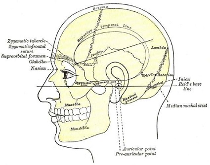
Picture 2.1 Sign Surface Anatomy Skull And Skull Frontal And Bone Zygomatic 15
Neuroanatomy
Facial paresthesia is a common sequelae of ZMC fractures given the proximity of sensory nerves, such as the infraorbital nerve, zygomaticofacial nerve, and zygomaticotemporal nerve (all branches of cranial nerve V2) to the zygoma.
Nerve infraorbital go out from jaw on through foramen infraorbital, in medial articulation between the maxilla and the zygoma. The infraorbital nerve provides sensory input from the cheek, upper lip, nose, and maxillary anterior teeth.
The zygomaticofacial and zygomaticotemporal nerves transmit sensory input from the lateral cheek and anterior temporal areas, respectively. They are branches of the zygomatic nerve that emerge in the pterygopalatine fossa and enter the orbit through the inferior orbital fissure before traveling along the lateral orbital wall. The zygomaticofacial and zygomaticotemporal branches then exit through foramina in the zygoma.
Severe ZMC fractures can also cause ipsilateral facial paralysis because the facial nerve is closely related to the zygomatic arch. Frontal branch of the facial nerve arises from the parotid glands deep into the superficial musculoaponeurotic system and crosses the zygomatic arch on its superficial surface. The branches frontal Then transition to surface lower fascia temporoparietal, where it runs to innervate the frontalis muscle 16,17
Anatomy Muscular 14
Muscle temporalis originate from throughout line temporal bone parietal And frontal and runs deep to the ZA to insert into the coronoid process of the mandible; it also has attachments to the zygoma. A depressed fracture of the zygomatic arch may impede movement of the temporalis muscle and cause trismus.
The masseter muscle originates from the inferior aspect of the zygoma and zygomatic arch and inserts at the angle of the mandible. It is a powerful muscle of mastication, and its contraction can displace an unstable segment of bone inferiorly in certain cases.
Zygomaticus major And minor is muscle expression face Which originate from the zygoma and inserted near the modiolus commissure of the mouth to aid elevation and lateralization of the corner of the mouth during smiling.
2 Classification Fracture Zygoma
Various system classification has used For categorize fracture ZMC. Below is the system proposed by Zingg et al in 1992:
Type A: fracture zygomatic No complete Which involving One articulation zygoma
A1: fracture arch zygomatic (ZMC)
A2: fracture wall orbital lateral
A3: rim fracture infraorbital
Type B: fourth articulation broken (fracture tetrapod complete), with the zygomatic bone itself remains intact
Type C: multi-fragment zygomatic fracture, where all four articulations are broken, and the body of the zygoma is also broken.
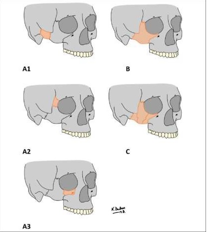
Picture 2.2 Classification Fracture Zygoma 18
3 Symptom Clinical
Symptom clinical fracture ZMC Which appear depends on pattern fracture and expansion of the fracture line. Clinical symptoms that can be found include: 12
Painful
Asymmetrical face
Ecchymosis And periorbital hematoma
Epistaxis
Deformity area rim infraorbital
Disturbance movement eyeball
Disturbance vision like diplopia, epiphora, or cornea dry
Enophthalmus or exophthalmos
Trismus
Dysfunction temporomandibular joint (TMJ)
Hypothesis, paresthesia, anesthesia And neuropraxia throughout distribution n. infraorbita
4 Diagnosis
Moment evaluate patient with broken bone zygoma, important For determine the mechanism and time of injury. Determine whether the injury was caused by trauma. blunt or trauma Translucency is also important, due to penetrating trauma more tends to involve deeper neurovascular structures. 19 A history of facial trauma or previous facial surgery may complicate fracture repair and should be ascertained preoperatively.
Diagnosis of zygomatic fractures begins with anamnesis and physical examination. Examination physique patient with trauma face Possible difficult Because existence edema and ecchymosis, making it important to perform a radiological examination. Plain Waters and submentovertex radiographs are used to diagnose zygomatic fractures, but Computed Tomography (CT) scanning is the gold standard that allows for a more detailed view. framework face two dimensions (2D) or three dimensions (3D). When fracture zygoma involving the orbit, CT scanning may be useful in determining the extent of the orbital fracture as well as findings such as entrapment, enophthalmos, or proptosis. 20,21
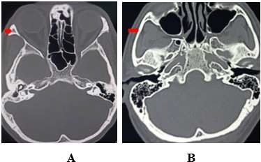
Picture 2.3 Overview CT scan A) Bone zygoma, B) Curved zygomatic (ZA) 15
5 Diagnosis Appeal
The zygoma contributes most of the inferior and lateral orbital walls. Any fracture of the zygoma requires evaluation for possible fracture or injury to the orbit, especially open globe injury. Cervical spine injury should be excluded, as should concomitant facial fractures, including sinus fractures. frontal, nose, face part middle (for example, Le Fort, naso-orbito-ethmoid), and broken bone mandible. Trauma part middle face can cause formation of a nasal septal hematoma, which should be assessed and managed to avoid septal necrosis and perforation or saddle nose deformity. Finally, malocclusion may result from mandibular or midface trauma and should be evaluated and managed as well. 1,6,22
6 Governance Fracture Zygoma
Various surgical approaches and treatment strategies have been proposed to achieve optimal treatment. successful approaches, including the temporal Gilles, coronal, brow, upper eyelid, transconjunctival, lower eyelid infraciliary, and intraoral vestibular approaches.5,6,22,23 An adequate surgical approach to reduce a fracture of the zygoma should provide the maximum exposure necessary to the segment. Which broken, minimize potential injury structure face, And ensure good functional and cosmetic results.
Management zygomatic fracture in a way line big can classified to be 3 category: management medical, reduction closed, And fixation internal reduction open.
Governance Medical Non- operative
Fracture zygoma usually can handled with observation just If displacement
minimal or no fracture segment. In addition, medical management may be an option if there are other comorbidities that prevent safe surgery. There is no strong evidence to support the use of prophylactic antibiotics for patients with upper and midface fractures. However, a number of expert surgery prescribe antibiotics during 5 until 7 days, especially if there is maxillary sinus involvement. If prescribed, antibiotics should include flora sinonasal, for example amoxicillin-clavulanate. 24 Besides That, diet Soft is usually recommended to prevent fracture disorders. bone due to muscle contractions.
Reduction with fixation external
Fixation external to fracture zygoma is technique Which when used in the appropriate clinical context, providing improved form and function with minimal exposure required.
First, the bone is palpated, and the fracture line is marked with an “X” on the outside of the skin. After injection of local anesthesia, fracture repair begins with approach Gillies with make incision throughout 1 centimeters in temporal hairline. After passing through the skin and subcutaneous tissue, the temporoparietal fascia and deep temporal fascia are found and then incised. Hemostat Kelly Which curved Then applied For facilitate dissection area superficial muscle temporalis in a way inferomedial going to arch zygomatic. Dissection is performed medially, or deep, to the zygomatic bone, and a uterine dilator is then inserted into the plane created by the Kelly hemostat. The use of a medium-sized dilator is recommended (Figure 2.4). 25
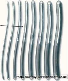
Picture 2.4 Dilator uterus used For reduction open fracture arch isolated zygomatic .
Sign arrow pointing to dilator size currently, Which recommended. 25
External force is applied under the zygomatic bone to lift the fragment and reduce segment Which broken, with attention Be careful so that No applying posterior pressure to prevent skull fracture. After confirmation of the improved contour of the malar eminence by external palpation, external fixation is initiated. Two sutures are placed around the zygomatic bone using 1-0 polypropylene suture with a curved and tapered needle to avoid facial nerve injury. After the 2 sutures are placed, an aluminum orthopedic plate with foam (Figure 2.5) is contoured to the unaffected contralateral zygomatic bone and secured in place using the sutures placed above. The plate should be placed (lateral-to-medial) from the pretragal to the infraorbital area midway between the lateral canthus and the nasal sidewall (Figure 2.6). It is important to trim the metal portion of the plate and leave the foam in place longer to prevent skin irritation and pressure necrosis. The plate remains in place for 3 weeks to allow for proper healing.
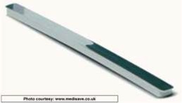
Picture 2.5 Plate orthopedics aluminum with foam used For fixation external. 25
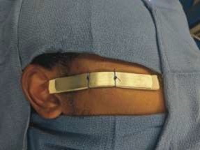
Picture 2.6 Position Which Correct from plate fixation external Which secured on the place with two 1-0 polypropylene sutures. 25
Reduction with fixation internal (ORIF)
Choice technique operative ORIF can done with various technique approach incision locations, including through scars, subciliary, transconjunctiva , coronal, temporal (Gillies), buccal, subtarsal, and so on.
Access fixation For reach frame face done on places certain considerations with consideration of aesthetic value in addition to ease of reaching it. To reach the anterior maxilla, an incision is made in the gingivobuccal sulcus/sublabial incision. The infraorbital rima, orbital floor, zygomaticofrontal area and upper maxilla can be reached through blepharoplasty or subciliary incision. For the frontal, nasoethmoidal, lateral orbital, zygomatic arch areas, a coronal incision is performed if necessary. 13,26 To reach the zygomaticomaxillary buttress, an incision can be made through incision sublabial. Incision sublabial his approach more easy but has the risk of damaging the infraorbital nerve. 26
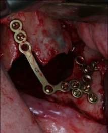
Picture 2.7 Fracture zygomaticofrontal exposed through laceration on skin Which expanded. 27
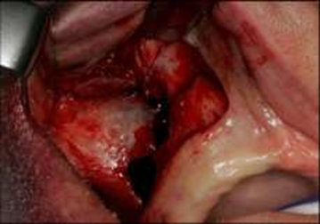
Picture 2.8 Fracture ZMC exposed through approach intraoral. 27
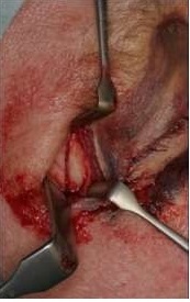
Picture 2.9 Rhyme infraorbital And floor orbit exposed through incision subciliary 27
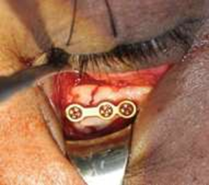
Picture 2.10 Approach mid-lid or subtarsal to edge infraorbital with fixation mini plate 28
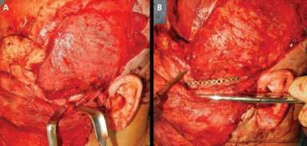
Picture 2.11 Approach coronal For reduction open And fixation internal arch comminuted zygomatic 28
The Gillies temporal approach has become a commonly used surgical technique for the reduction of zygomatic complex fractures. However, this surgical approach is associated with used wound face in line hair And risk paralysis facial nerve. Especially exposure more carry on from zygomaticofrontal or edge orbital inferior is required for placement of mini-plate fixation in case of unstable zygomatic complex fractures. Surgical reduction of zygomatic fractures with a surgical approach intraoral First time explained on 1909 by Keen 28 , And a number of
study Then documenting results treatment after reduction open zygomatic complex fractures with an intraoral surgical approach.
Currently, internal fixation with small plates and screws is preferred because it is easier and more practical. Mini plates using 2 mm screws are used for maxillary buttress stabilization. Such a small size is used so that the plate is not visible and palpable. Mini plates are used so that they can passively adapt to the desired frame contour. Drilling to install the screws is done with a sharp drill bit with the right diameter. Previously, the screws were cooled to avoid bone dermal necrosis and were carried out at a low drilling speed. Titanium plates and screws are the gold standard used to maintain the stability of the repositioned fracture segment. However, its use requires a second operation to remove it and its use in children can inhibit bone growth where the bone may grow around the plate resulting in bone deformity. Another alternative is an absorbable plate that does not require a second operation to remove the plate, this plate can maintain his strength until absorbed. On children No inhibits facial bone growth and causes less pain. 13,29,30
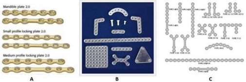
Picture 2.9 Types mini plate. A) Titanium mini plate, B) Absorbable mini plate,
C) Types form miniplate. 13.30
Algorithm management based on system classification fracture Zingg: 2
Type A1 (zygomatic arch fracture): Non-displaced ZA fractures are often managed conservatively. In contrast, displaced ZA fractures require reduction, which can be performed via the Gillies approach with use incision temporal, screw Carroll Girard transcutaneous directly in on location fracture Which stressed, or approach Keen through incision intraorally in the maxillary vestibule. Isolated arch fracture that is unstable after reduction can splinted in a way external with wire steel or stitching thick or
coated through incision coronal. 8
Type A2 (lateral orbital wall fracture): Reduction and fixation of this area is best accomplished through a lateral brow incision or an upper blepharoplasty incision. Miniplates provide adequate stabilization of these fractures.
Type A3 (infraorbital rim): The infraorbital rim should be reduced and lined to return its normal contour and done approach through a transconjunctival or subciliary lower eyelid incision.
Type B (tetrapod fracture): Mildly displaced tetrapod fractures can sometimes be reduced by the Gillies or Keene approach. Most tetrapod fractures, however, remain unstable after attempts at reduction. the And on Finally need reduction open And fixation internal with single-point fixation (zygomaticomaxillary or zygomaticofrontal suture), fixation 2 point (suture ZM And ZF), or fixation 3 point (ZM). and sutures ZF, And infraorbital edge). Final, ZA can coated through incision coronal or laceration pre-existing as a fourth fixation point if necessary.
Type C (fracture tetrapods comminuted): Fracture This is indication absolute for open reduction and internal fixation (ORIF). Treatment is similar to that used For fracture type B, with ORIF on support ZM, edge infraorbital, ZF suture, zygomaticotemporal suture, and zygoma fragment if necessary.
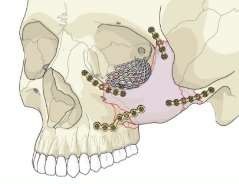
Picture 2.9 Fixation 4 point with miniplate . 20
8 Complications
Complications from broken bone zygoma can relate with injury beginning or operative management and may include:
Painful
Infection wound
Facial asymmetry
Network scar
Bleeding (epistaxis)
Failure device hard (exposure, touch)
Paresthesia nerve infraorbital
Sensitivity temperature
paresis or facial paralysis
Results bad cosmetics
Trismus 5.31
Orbital complications (usually related to concomitant orbital floor fractures ):
Blindness
Decrease sharpness vision
Malposition ectropion/entropion/eyelid eye
Exposure/abrasion cornea
Ptosis
Epiphora
Enophthalmos/dystopia orbital
Diplopia
Syndrome orbital fissure superior
Complications of zygomatic complex fractures can occur due to initial trauma, surgical intervention, or inaccurate surgical treatment. It has been reported that up to 5.5% of patients require a second procedure for zygomatic complex fractures within 4 weeks of initial repair due to inadequate reduction. 31
9 Maintenance And Rehabilitation Post Operation
Maintenance And recovery post operation varies depends on level injury and the reconstruction method used. Regardless of the severity of the fracture or the technique chosen, patients should be advised to refrain from strenuous activity for at least 2 weeks to allow for complete healing with minimal bruising and swelling. Depending on the fracture involved, additional postoperative care may include: 15
Lubricants drops eye (broken) bone orbital)
Nasal irrigation
Drug gargle (when incision intraoral use)
Diet soft (fracture ZA displaced or fracture mandible/malocclusion)
The skin incision line requires application of antibiotic ointment (must be an eye ointment). For incision periorbital) at least 72 O'clock post operation before switch to petrolatum ointment until the incision is completely healed. Avoiding sun exposure and proper sun protection, as well as using silicone-based scar creams and lotions can help improve the appearance of scars. Postoperatively, patients should be closely monitored for potential complications, especially Which relate with infection And complaint vision. Visit usually performed 1 week after surgery and then every few weeks thereafter until the fracture appears stable and any complications that may arise have been resolved. 15
10 Consultation
Every patient with trauma who Enough to maintain fracture ZA or ZMC should undergo a full trauma evaluation to rule out any other injuries. simultaneously For determine consultation Which most appropriate (for example, surgery neurology, ophthalmology, orthopedic surgery, plastic surgery, ENT, oral-maxillofacial surgery, vascular surgery). In addition to primary craniofacial trauma surgery services (otolaryngology/oral-maxillofacial surgery/surgery plastic), consultant Which most Possible is doctor eye or oculoplastic surgeons due to the high rate of ocular injuries accompanying ZMC fractures.
Injury eye accompanying Which most general is bleeding subconjunctiva, which occurs in 67% to 87% of patients with ZMC fractures. Other less common ocular complications include symptoms of diplopia (16% of patients), hyphema (13%), decreased visual acuity vision (13%), traumatic optic neuropathy (6%), choroidal rupture (6%), corneal abrasion (5%) , bleeding vitreous (3%), ablation retina (3%), bleeding retina (1.5%), And
mydriasis (1.5%). 32.33
11 Prognosis
Comminuted fractures in ZMC have worse outcomes than comminuted fractures. non-comminuted, with level operation corrective 33% more height 31 A number of (5%-9%) patients, including those with non-comminuted fractures, require a second operation due to inadequate fracture reduction. 27,31 Indications for revision surgery include persistent enophthalmos, diplopia, facial contour irregularities, and facial dysfunction. temporomandibular joint. 20 Some degree of postoperative asymmetry occurs in 20% to 40% of patients, with major asymmetry occurring in 3% to 4%. Paresthesia may persist long-term in 22% to 65% of patients. 34
CONCLUSION
Zygoma is support main framework face part lateral middle third that provides normal cheek contour. The main etiology of zygoma fracture is traffic accident. Diagnosis of zygoma fracture begins with anamnesis, physical examination, and supporting examination. Computed Tomography (CT) is the gold standard supporting examination to establish the diagnosis of zygoma fracture. Zygoma fracture is most often treated with open reduction internal fixation. Inappropriate reduction of zygoma fracture can cause facial asymmetry and malposition, so it is important to be aware of this case from anamnesis to find the etiology and mechanism of trauma, physical examination, and diagnosis. The decision and type of surgical treatment of zygoma fracture depends on several issues that need to be considered based on the case.
REFERENCE
Bogusiak K, Arkuszewski P. Characteristics and epidemiology of zygomaticomaxillary complex fractures. J Craniofac Surg. 2010;21(4):1018–1023.
Marine R Oh, Freire-Maia B. Management of fracture of the zygomaticomaxillary complex. Oral Maxillofac Surg Clin North Am. 2013;25(4):617–636.
Fonseca R J, Barber H D, Powers M P, Frost D E. Saint Louis, MO: Elsevier Health Sciences; 2013. Oral and Maxillofacial Trauma; pp. 354–450.
Louis M, Agrawal N, Kaufman M, Truong T A. Midface Fractures I. Semin Plast Surg. 2017;31(02):85–93.
Birgfeld C B, Mundinger G S, Gruss J S. Evidence Based Medicine: Evaluation and Treatment of Zygoma Fractures. Plast Reconstr Surg. 2017;139(01):168e–180e.
Ellis E, III, Reddy L. Status of the internal orbit after reduction of zygomaticomaxillary complex fractures. J Oral Maxillofac Surg. 2004;62(03):275– 283.
Choi K Y, Ryu D W, Which J D, Chung H Y, Cho B C. Feasibility of 4-point fixation using the preauricular approach in a zygomaticomaxillary complex fracture. J Craniofac Surg. 2013;24(2):557–562.
Hindi IN, Mutterties CE, Mehta C, Boukovalas S, Lee JC, Bradley JP. Treatment of isolated zygomatic arch fracture: improved outcomes with external splinting. Plastic Reconstr Surg. 2017;139(5):1162e–1171e.
Evans GR, Daniels M, Hewell L. An evidence-based approach to zygomatic fractures. Plast Reconstr Surg. 2011;127(2):891–897.
Orabona GD, Abbate V, Maglitto F, et al. Postoperative management of zygomatic arch fractures: in-house rapid prototyping system for the manufacture of protective facial shields. J Craniofac Surg. 2019;30(7):2057–2060.
Liu, F.C., Halsey, J.N., Oleck, N.C., Lee, E.S., & Granick, M.S. (2019). Facial Fractures US a Results of Falls in the Elderly: Accompanying Person Injuries and Management Strategies. Craniomaxillofacial Trauma & Reconstruction, 12, 45 - 53.
Donald PJ, Sykes J. Facial Fractures. In: Ballenger's Otorhinolaryngology Head and Neck Surgery. 16th Edition. London: Hamilton. 2003;39:p.900-50.
Act S, Gonul Oh, Satilmis T, Garip H, Goker K. Management of midfacial fracture. In: A Textbook of Advanced Oral and Maxillofacial Surgery. Intech. 2013; 416-45.
Alcala AG, Arribas IJ, Martin MA, Romance A, Montalvo JJ, Milan JM. Pediatric Facial Fractures: Children Are Not Just Small Adults. Radiographics. 2008; 28:441-61.
Bergeron JM, Raggio BS. Zygomatic Arch Fracture. [Updated 2024 Jan 26]. In: StatPearls [Internet]. Treasure Island (FL): StatPearls Publishing; 2024 Jan-. Available from: https:// www.ncbi.nlm.nih.gov/books/NBK549898/
Lee EI, Mohan K, Koshy JC, Hollier LH. Optimizing the surgical management of zygomaticomaxillary complex fractures. Seminole Plastic Surg. 2010 Nov;24(4):389-97.
Agarwal CA, Mendenhall SD, Foreman KB, Owsley JQ. The course of the frontal branch of the facial nerve in relation to the fascial planes: an anatomical study. Plast Reconstr Surg. 2010 Feb;125(2):532-537.
Zingg M, Laedrach K, Chen J. Classification and treatment of Zygomatic fractures: a review of 1,025 cases. J Oral Maxillofac Surg. 1992;50(08):778–790.
Strong EB, Gary C. Management of Zygomaticomaxillary Complex Fractures. Facial Plast Surg Clin North Am. 2017 Nov;25(4):547-562.
Balakrishnan K, Ebenezer V, Dakir A, Kumar S, Prakash D. Management of tripod fractures (zygomaticomaxillary complex) 1 point and 2 point fixations: A 5-year review. J Pharm Bioallied Sci. 2015;7(Suppl 1):S242-S247. doi:10.4103/0975- 7406.155937
Peretti N, MacLeod S. Zygomaticomaxillary complex fractures: diagnosis and treatment. Curr Opin Otolaryngol Head Neck Surg. 2017 Aug;25(4):314-319. doi: 10.1097/MOO.0000000000000372. PMID: 28548998.
Farber SJ, Nguyen DC, Skolnick GB, Woo AS, Patel KB Current management of zygomaticomaxillary complex fractures: A multidisciplinary survey and literature review. Craniomaxillofac. Trauma Reconstruction. 2016;9(4):313–322. doi: 10.1055/s-0036-1592093.
Ellstrom CL, Evans GR Evidence-based medicine: Zygoma fractures. Plast. Reconstr. Surg. 2013;132(6):1649–1657. doi: 10.1097/PRS.0b013e3182a80819.
Cohn JE, Othman S, Bosco S, et al. Management of Isolated Zygomatic Arch Fractures and a Reviews of External Fixation Techniques. Craniomaxillofac Trauma Reconstr. 2020;13(1):38-44. doi:10.1177/1943387520905164
Hopper RA, Salemy S, Sze RW. Diagnosis of Midface Fractures with CT : What the Surgeon Needs to Know. Radiographics. 2006; 26:783-93.
Starch Jensen T, The Linnebjerg LB, Jensen JD. Treatment of Zygomatic Complex
Fractures with Surgical or Nonsurgical Intervention: A Retrospective Study. Open Dent J. 2018 May 21;12:377-387. doi: 10.2174/1874210601812010377. PMID: 30202484; PMCID: PMC6129990
27 Money HD, Dillon J. Contemporary Management of Zygomaticomaxillary Complex Fractures. Semin Plast Surg. 2021;35(4):256-262. Published 2021 Oct 7. doi:10.1055/s-0041-1735812
28. Lore JM, Klotch DW. Fractures of facial Bones. In: An Atlas of Head & Neck Surgery. 4th ed. Elsevier Saunders. 2005; 13: p.595-650.
29. Maxillofacial Reconstruction Plastic Code Perhati-KL. Workshop Management of Maxillofacial Fracture. Semarang: 2015; 2-71.
30. van Hout WM, Van Cann EM, Koole R, Rosenberg AJ. Surgical treatment of unilateral zygomaticomaxillary complex fractures: A 7-year observational study assessing treatment outcome in 153 cases. J Craniomaxillofac Surg. 2016 Nov;44(11):1859-1865.
31 Jamal BT, Pfahler SM, Lane KA, Bilyk JR, Pribitkin EA, Diecidue RJ, Taub DI. Ophthalmic injuries in patients with zygomaticomaxillary complex fractures requiring surgical repair. J Oral Maxillofac Surg. 2009 May;67(5):986-9.
32. Malik AH, Shah A A, Ahmad I, Shah BA. Ocular Injuries in Patients of Zygomatico- Complex (ZMC) Fractures. J Maxillofac Oral Surg. 2017 Jun;16(2):243-247.
33. Rajantie H, Snäll J, Thorén H. Postoperative temporomandibular dysfunction in patients with fracture of the zygomatic complex: a prospective follow up study. Br J Oral Maxillofac Surg. 2018 Sep
Comments