Diagnosis And Treatment Auricular Keloid
I Made Nudi Arthana
Auricular Keloid is a pathological wound healing process due to the fibroproliferative process of abnormal cutaneous tissue where fibroblastic proliferation is not balanced with the apoptosis process. Keloid marked with existence of network growing scar beyond the limits of the original wound area and this scar tissue rarely undergoes spontaneous regression. Trauma, inflammation, surgical scars, and burns can cause scars where if the healing of this wound occurs excessively it will cause the formation of keloids. Although it does not cause mortality, keloids can affect the quality life in a way significant Because influence cosmetics And cause a feeling uncomfortable due to taste painful or itchy that accompanies it. By Therefore, proper keloid management is very important. 1,2
LITERATURE REVIEW
Anatomy And Histology Skin
The skin is the largest organ in the human body. Anatomically, the skin is composed of three layers, namely the epidermis, dermis, and hypodermis, where these three layers of skin have significant variations in terms of structure and function. 5,6
Epidermis
The epidermis is divided into the stratum basale (the deepest part of the epidermis), stratum spinosum, stratum granulosum, stratum lucidum, and stratum corneum (the outermost part of the epidermis). The stratum basale separates the epidermis from the dermis and is attached to the basement membrane via hemodesmosomes. The stratum basale is composed of cuboidal epithelial cells to columnar Which active to split to form cell keratinocytes. Besides that, on stratum basal Also found cell melanocytes Which act in skin pigment synthesis. The stratum spinosum is composed of 8-10 layers of keratinocyte cells where adjacent cells are bound to each other through desmosomes. In this layer, dendritic cells are also found which have an immune function. Stratum granulosum arranged on 3-5 layer cell shaped diamond with granule keratohyalin and lamella. Stratum lucidum consists of 2-3 layers of keratinocyte cells which are generally found in the deeper parts of the skin. thick like on area palm hand And palm foot. Layer The stratum lucidum contains eleidin which is a product of keratohyalin. The last layer is the stratum corneum which is the thickest layer consisting of from 20-30 layers of cells dead keratinocytes (cell flat without nucleus). Thick layer from These dead keratinocytes are the first form of immune defense of the human body. 5,6
Dermis
The dermis layer is the layer that is located directly below the epidermis layer separated by the basal membrane. The dermis consists of 2 layers, namely the papillary layer and the reticular layer. The papillary layer is the layer of the dermis that borders directly on the epidermis. This papillary layer is thin and consists of loose connective tissue. The reticular layer is a deeper layer of the dermis, contains fewer cells, but is composed of dense connective tissue/collagen fibers. Sweat glands, hair, hair follicles, muscles, sensory nerves, and blood vessels are also found in the dermis. 5,6
Hypodermic
The hypodermis is the deepest layer or also known as fascia. subcutaneous. Layer This contain cell adipocytes, follicle hair, sensory receptors, nerves, and blood vessels. 5,6
Anatomically, the skin is a highly vascular organ. The supply skin blood generally comes from from plexus of blood vessels found between layer reticularis And layer papillary on dermis. Besides the plexus blood vessels, lymphatic tissue is also found between the reticular layer and the papillary layer of the dermis. Nerves on the skin generally consists of from nerve somatic and autonomic nervous system. The somatic sensory nervous system generally plays a role in the process of receiving sensory stimuli by special cutaneous sensory receptors, such as receptors Merkel (For touch light), corpus Pacinian (For pressure in and vibration), Meissner corpuscles (for light pressure), Ruffini corpuscles (for deep pressure and stretch), and free nerve endings (pain). These receptors will then change stimuli from the external environment into nerve stimuli which will then be transmitted further through the sensory nerve pathways to the spinal cord according to the dermatome of skin innervation. 5,6

Picture 1. Structure Anatomical And Histology Skin Network . 6
Anatomy Auricular Lobe
The auricular lobe is part of the external ear located in the inferior from pinna (auricular). Slightly different with structure Pinna, the auricular lobe is composed of skin and fatty tissue without cartilage. The auricular lobe has many nerves. and blood vessels. Although the auricular lobule is not own function specific, lobule auricular own function aesthetic And erogenous function . 7
Supply blood on pinna done by artery temporalis superficial And posterior auricular artery which then forms perforating arteries in the helical rim and auricular lobule. Vascular drainage is carried out by a combination of superficial temporal veins, posterior auricular veins, and mastoid emissary veins. The pinna as a whole is innervated by branches of cranial nerves V, VII, IX and X. 8,9

Picture 2. Anatomy Auricular Lobules on Pinna 7

Picture 3. Supply Vascular on Pinna. 10
Definition Keloid
Keloid is a form of pathological scar tissue formation. Term keloid First time known in literature modern on beginning 19th century by Jean Louis Aliber who is the father of French Dermatology. Keloids were first described as benign skin tumors that have a growth horizontal invasive exceed limit wound until invade skin around. 11.12
Histologically, keloids can be defined as a form of abnormal wound healing characterized by excessive fibroproliferation composed of irregular collagen type I and III fibers. In contrast to hypertrophic scars, keloids are characterized by excessive fibroproliferation composed of irregular collagen type I and III fibers. ( hypertrophic scar ), keloid generally level severity more heavy and have size that exceeds the wound boundaries. Some differences between keloids and hypertrophic scars are shown in Table 2.1. 4,11
Table 2.1 Differences Keloid And Hypertrophic Scars . 11
Characteristics | Keloid | Hypertorphic Scars |
Growth | Beyond limit woundoriginal | No exceed limitwound original |
Histology | Composed on fiber collagen typeI And III randomly arranged | Composed of collagen type fibers III Whicharranged parallel |
Prognosis | Almost No Once healed spontaneously | Can healed in a way spontaneous in several years |
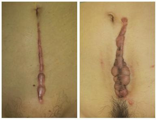
Picture 3. Difference Keloid And Hypertrophic Scars . 13
Epidemiology Keloid
Incident keloid varies And very influenced by ethnicity And race. Risk The incidence of keloids increases with increasing skin pigmentation. The incidence of keloids in Black and Hispanic populations varies from 4.5% to 16%. The incidence of keloids in Asian populations in general is estimated to be 0.2% to 5.2%, while the incidence of keloids in Caucasian populations is reported to be around <1%. Although skin pigmentation is considered a determining factor that differentiates variations incident keloid on race Which different, reported that incident keloid in the African population with albinism around 7.5% where this is no different from the incident keloids in the population Africa in general namely around 8.3%. By Therefore, it is suspected that there are also genetic factors that cause differences in keloid incidence between races and ethnicities. 2,12
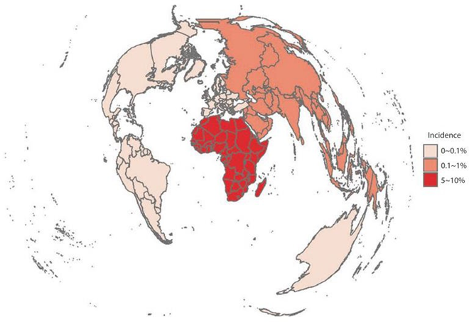
Picture 4. Map Distribution Incident Keloid. 13
Keloid incidents have also been reported to be more common in the first to third decade of life. Other studies have reported the same thing where the prevalence keloid highest found on population age 10-29 year. However, There isn't any difference significant between man And Woman in incident keloid. The thing that The same Also reported by studies Liu et al. Which involving population students in China. Liu et al. showed that the prevalence of keloids did not differ significantly between man And Woman. On the contrary, studies Manohara et al. show that the incidence of keloids was higher in male patients with a male to female ratio of 3:2. This is thought to be because men experience keloids more often. trauma. However, Shaheen et al. actually stated that the incident keloid more tall on Woman Because population Woman more often undergo ear piercing which is the main cause of keloids, especially keloids in the auricular lobe. 2,14–16 The distribution of keloids is found most in the upper extremity area with the most common cause being trauma. This has also been reported by studies Manohara et al. Which show etiology keloid mainly caused by trauma (50%), followed by scars due to acne (15%), surgery (10%), infection herpes shingles (10%), wound burn (5%), And happen spontaneously (10%). 2,14,15
Etiology And Factor Risk Keloid
Keloids can occur due to trauma, inflammation, scars from surgery, and burns can cause wounds to the skin tissue. Keloids are considered as a the disease that is multifactorial. There is a number of factor which is thought to increase the risk of keloid development in certain populations.
Factor genetics
Patients with a history of keloids in family members have a higher risk of keloids, so it is suspected that keloids are closely related to certain genetic predispositions. The inheritance pattern of this genetic predisposition is not yet clear. known Certain, but allegedly nature autosomal recessive or autosomal dominant with penetration incomplete. There is a number of mutation genetics which is thought to be responsible for the predisposition to keloids in certain populations, but no single gene mutation has been proven to date. Several genetic mutations have been shown to be associated with genetic predisposition from the occurrence keloid is Single Nucleotide Polymorphism (SNP) at loci 1q41, 3q22.3-23, 15q21.3, 2q23, and 7p11. In addition, other studies show existence connection between status HLA-DRB1*15 with the risk of keloids in the white-skinned population. Individuals who are carrier HLA-DQA1*0104, DQB1*0501, And DBQ*0503 Also have been found to have an increased risk of keloid formation after injury. Certain syndromes associated with abnormal connective tissue mutations have also been reported to increase the risk of keloid formation, such as Rubinstein-Taybi syndrome (RSTS), Ehlers-Danlos syndrome, Lowe syndrome, and X-linked syndromes . 13,16
Factor systemic (hormonal and immunity)
Keloid incidents are said to occur more often at a young age but not in the first decade of life. In addition, keloid incidents are also more often found in pregnancy and puberty, so it is suspected that there is a role for sexual hormones that influence keloid growth.
Factor environment
Environmental factors also contribute to the occurrence of keloids. The presence of environmental factors trauma due to objects sharp (including piercing ear) is the most common cause of keloids. In addition, keloids most often occur in areas of skin that have high tension or friction (such as the chest, scapula, lower jaw, neck, upper extremities, lower abdomen, femur, and knees). Areas of skin that have Mechanical tension ( skin tension ) is thought to accelerate keloid growth. 13,16
Pathophysiology Keloid
wound healing is greatly influenced by the role of fibroblasts. Normal fibroblast activity is needed for the production of growth factors and cytokines which then trigger collagen production. Keloid formation is the result of an imbalance between increased synthesis and decreased degradation of collagen synthesis and extracellular matrix. Improvement synthesis collagen And matrix extracellular by fibroblasts are caused by overexpression of the pro-inflammatory mediator TGF-β1. Expression excessive from TGF-β1 And TGF-β2 will cause decline TGF-β3 production which causes increased fibroblast activity and the formation of collagen synthesis and extracellular matrix. In addition, increased expression of TGF-β1 will cause improvement tissue inhibitors of metalloproteinases (TIMP) And decline production matrix metalloproteinase (MMP). TEAM And MMP plays a role important in process degradation collagen And matrix extracellular. Thus, the increase in TIMP and decrease in MMP will cause a decrease in collagen and extracellular matrix degradation. In addition to TGF-β expression, the expression of protein pro-inflammatory other, like Vascular Endothelial Growth Factor (VEGF) And Platelet-Derived Growth Factor (PDGF), Also contribute against the overproduction of collagen. VEGF and PDGF activity will increase the activity fibroblasts consequence activation track mechano-transduction Which stimulated by stress Mechanic consequence voltage skin on area body certain, like sternum, shoulder, and suprapubis. This is because fibroblasts are also sensitive to mechanical tension of the skin so that their activity is influenced by skin tension. Increased mechanical tension will trigger the differentiation of fibroblasts into myofibroblasts which will cause increased expression of TGF-β1. 11,13,17

Picture 5. Scheme Illustration Process Healing Wound ( Wound Healing ) Normal and Pathological in Keloid Formation. 13
The process of keloid formation in the auricular lobule after trauma or ear piercing goes through the same process. Trauma to the skin area or subcutaneous tissue of the auricular lobule will trigger the wound healing process in the auricular area. The presence of an excessive inflammatory response in susceptible individuals (either due to genetic or hormonal influences) will trigger the formation of keloids. 18
Diagnosis Keloid
Clinical diagnosis of keloids can be done easily through anamnesis and simple physical examination. Supporting examinations are generally not needed. required. Anamnesis generally required For know existence risk factors and history of injury or trauma to the area of keloid growth. Simple physical examination (visual inspection and palpation) of the skin is sufficient to direct a subjective diagnosis of abnormal scar tissue formation. Clinically, keloids are distinguished from hypertrophic scars based on their appearance. Keloids generally grow exceed limit wound original, whereas hypertrophic scars do not grow beyond the boundaries of the original wound (Figure 3). In addition, keloids are generally accompanied by elevation, hyperemia, and have a more abnormal shape than hypertrophic scars. Some patients may also experience symptoms of itching and discomfort in keloid tissue, but this is not found in patients with hypertrophic scars . 16,19
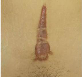
Picture 6. Lesion Intermediate Which Difficult Differentiated.
Although difference simple is able to differentiate almost all
In cases of keloids and hypertrophic scars , some cases of keloids and hypertrophic scars may be difficult to distinguish (Figure 6). Therefore, a rating scale is needed that can used For diagnose keloid in a way more objective. Wrong One example score objective Which can used For help differentiate diagnosis keloid and hypertrophic scar objectively is the Japan Scar Workshop Scar Scale (JSS) 2015. JSW scores range from 0 to 25 where JSW scores of 0-5 lead to a diagnosis of mature scars , JSW scores of 6-15 lead to a diagnosis of hypertrophic scars, and JSW scores of 16-25 lead to a diagnosis of keloids. 20 In addition to the JSS 2015, several other assessment scales that can be used are The Vancouver Scar Scale (VSS), Manchester Scar Scale (MSS), Patient and Observer Scar Assessment Scale (POSAS), Visual Analog Scale (VAS), and Stony Brook Scar Evaluation Scale (SBSES). 19

Figure 7. Japan Scar Workshop Scar Scale (JSS) 2015. 20 Supporting examinations that can be performed to assist in the diagnosis
diagnosis keloids. Inspection support This especially needed if suspected tumor as a differential diagnosis of keloids. Some benign tumors that may have an appearance resembling keloids and hypertrophic scars include pseudolymphoma, mixed skin tumors, xanthogranuloma, leiomyoma, and dermatofibroma (Figure 8). Some malignant tumors of the skin can also resemble keloids and hypertrophic scars, such as dermatofibrosarcoma protuberans (DFSP), squamous cell carcinoma (SCC), and malignant melanoma (Figure 9). Diagnostic examinations that can be performed include ultrasonography, Computed Tomography (CT) -scan , or Magnetic Resonance Imaging (MRI). Ultrasonography (ultrasonic elastography) is preferred for differentiate keloid And hypertrophic scar from tumor. Elastography Ultrasound examination of keloids and hypertrophic scars will show areas with a hard consistency and lower echogenicity compared to the surrounding dermis, but the interior of the lesion can sometimes be heterogeneous. Ultrasonography with color doppler can help in evaluation of vascularization, thickness, volume, presence of blood flow in keloids indicating the type of vascularization, thickness of blood vessels, peak systolic velocity of blood vessels. Ultrasonography examination with color doppler can also be used to detect calcification or fistula to determine which therapy may be suitable for each keloid case and to assess the response of keloid scars to different modalities. 19,20
Histological examination is generally performed on tissue samples obtained during tissue resection or biopsy (especially if a tumor is suspected). tumor). Inspection histological keloid generally indicates arrangement collagen fiber type I And III Which No regular with infiltration cell inflammation And high fibroblasts And active mesenchymal cells excessively. Besides collagen, in keloid tissue, deposits of elastin, fibronectin, and proteoglycans can generally be found. In addition, thickening of the epidermis layer was found in keloid tissue. This thickening of keloid tissue is thought to occur due to abnormal differentiation of keratinocytes. 12,21

Picture 8. Tumor Benign Which Similar Keloid. a) Pseudolymphoma And b) Xanthogranuloma. 20
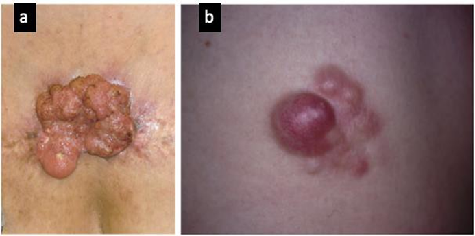
Picture 9. Tumor Malignant Which Similar Keloids. a) Dermatofibrosarcoma protuberans and b) Squamous cell carcinoma. 20
Type And Location Keloid
Allegedly there is connection between location anatomical And risk the occurrence keloid. This assumption is based on several phenomena where individuals who are treated experience keloids after wounds, but these keloids only occur in wounds in the area body certain. Until At the moment, there is a number of studies Which observe the role of anatomical location in keloid formation. Areas of keloid predilection include the anterior chest, shoulder, arm on, neck, And cheek. Area body other, like petals eye, palm hand, mucosa, genitals, And palm foot is area Which Keloids are rare. Keloids are more likely to occur in mobile areas with high skin tension, such as the shoulders, neck, and upper arms. Wang et al.'s study also showed that 72.0% of keloid cases in the neck and head area occurred in the ears. However, the auricular lobe is an area that is tension-free, but area This become predilection keloid so that Still The exact mechanism behind the anatomical location of keloid predilection is not yet known. 12,16,22
In general line big, there is two phenotype keloids. Keloid with type spreading “ spreading ” well in a way vertical and also horizontal marked with existence Keloids are irregular in shape with thickening mainly found in the edge area. and flatter in the central area that does not move. This type of keloid can be hypopigmented or hyperpigmented where hypopigmentation is more common. on area central And hyperpigmentation more frequently found on edge area. Another keloid edge is the “ bulging ” or raised type. Unlike the “ spreading ” type of keloid, this type of keloid has almost no flat area. 12
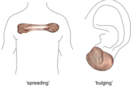
Picture 10. Type Keloid
Treatment Keloid
To date, there is no single therapy that is best or effective for all keloids. The location, size, and depth of the lesion and patient characteristics will be factors in determining the combination of therapies used. There is a number of choice therapy keloid Where choice therapy This generally combined For get external best: 11,16,19
Excision
Excision is Wrong One method Which most fast For remove large keloid lesions. However, this therapy has a high recurrence rate. so it must be combined with other therapies. Things to note when performing excision on keloids is to ensure that the edges of the sutures have minimal skin tension so as not to trigger the formation of new keloid tissue. This therapeutic modality is generally combined with other postoperative adjuvant therapies, such as radiotherapy, intralesional corticosteroid therapy, interferon (IFN) injection, compressive therapy, and others.
Cryotherapy
Cryotherapy is method surgery Which use Liquid nitrogen will cause cell and microvascular damage through intracellular crystallization, causing tissue anoxia. Generally, 1-3 freeze-thaw cycles are performed for 10-30 seconds for each lesion to achieve effect Which desired. Therapy can repeated every 20-30 day (every 3-4 weeks). The success rate of this therapy is estimated at 30-75%.
Injection intralesional corticosteroids
Corticosteroids can reduce collagen synthesis, production cytokines pro-inflammatory, and fibroblast proliferation during wound healing. To date, the most widely used corticosteroid as an intralesional injection modality is triamcinolone acetonide (TAC) with a concentration of 10-40 mg/mL. TAC is administered intralesionally with an interval of 4-6 weeks between injections. There is a studies which compares the efficacy injection TAC at various different doses where the results showed that TAC injection with dose of 20 mg/mL and 40 mg/mL provided comparable outcomes, but TAC injection at a dose of 10 mg/mL did not provide any improvement, so therapy with dose This stopped on studies. Injection TAC often given as therapy adjuvan post surgery. When used as therapy single, therapy This own level recurrence 33% on 1 year First And 50% on 6 year First.
Dressing occlusive And therapy compression
Dressing occlusive And therapy Compressive generally more nature preventive so that it is more widely used as Occlusive dressings used as keloid therapy include silicone gel sheeting (SGS), non-silicone occlusive dressings , and cordran tape . SGS is the most widely used type of occlusive dressing today. The antikeloid effect of this occlusive dressing combines occlusion and hydration to modulate mechanoreceptors that play a role in the process of apoptosis and decreased production of extracellular matrix. Dressing occlusive This at least must used during 12 hours per day for 12 months to achieve the desired effect. Compressive therapy also utilizes this theory where the pressure applied will have a thinning effect on the skin, thereby reducing the cohesion of collagen fibers in the wound or keloid tissue. This compressive therapy can use various compression modalities, such as elastic wrap , special compression molds, special earrings, bandages, or even magnets. Compression therapy is the best choice for auricular lobular keloids where the use of compressive therapy with a pressure of 24 mmHg for at least 12 hours per day for at least 6 months shows an effectiveness of 70-95. Compression pressure No may exceed 30 mmHg Because can cause tissue necrosis .
Radiotherapy
Laser
Radiation can destroy fibroblasts in wounds or scars, thereby reducing or preventing the formation of neovascularization. fibroblasts This will more continue to lower production collagen in the wound healing process.
Ablative laser therapy has The principle is almost the same as radiation that aims to destroy fibroblast cells. Ablative lasers include 10,600nm carbon dioxide laser, Erbium: Yttrium Aluminum Garnet (Er: YAG Laser) 1064nm, And laser Argon 488-nm. Laser non-ablative (like laserpulsed dye laser 585 nm) Work with produce process photothermolysis which will cause microvascular thrombosis thereby causing apoptosis cell fibroblasts. Therapy laser mentioned most effective used in patients with keloid duration of less than 2 years.
Agent topical or injection intralesional other
Several topical agents, such as 5-fluorouracil (5-FU), bleomycin, Imiquimod 5%, and tacrolimus, are also used as adjuvant therapy for keloids. 5-fluorouracil (5-FU) and bleomycin work as neoplastic agents that can inhibit DNA synthesis and cell proliferation, including fibroblast cells, without causing tissue necrosis. Imiquimod 5%, and tacrolimus are immunomodulatory agents that are expected to be able to apoptosis matrix extracellular And hinder production collagen. Besides In addition, heparin gel and flavonoids are also used because they have anti-inflammatory effects. Where decline inflammation trusted will increase MMP expression resulting in increased collagen degradation. Intralesional injection agents besides corticosteroids on keloid very much diverse. Verapamil, which is a calcium channel blocker (CCB), can inhibit interleukin-6 (IL-6) and VEGF, thereby reducing the production of extracellular matrix in keloids. Verapamil is generally given in the form of intralesional injection at a dose of 2.5 mg/mL. Botolinum toxin A (BTX-A). is generally injected into keloids that have high skin tension. BTX-A is injected intralesionally at a dose of 70-140 U with an interval of 1 or 3 months for 3 sessions.
Therapy other
There are several potential therapies that are still being researched today as governance keloids. Inhibitor VEGF (Bevacizumab), phototherapy, UVA-1 therapy, therapy narrow UVB band , TNF-α inhibitor (etanercept), Recombinant human interleukin (rhIL-10), or Human recombinant TGF-β3/TGF-β1 is considered as therapy potential which is able to reduce synthesis collagen in keloids.
As previously mentioned, there is no single therapy that is effective as a keloid therapy. Therefore, almost all literature uses a combination of therapies. Excision or cryotherapy of keloid tissue is generally followed by adjuvant therapy such as intralesional corticosteroid injection, laser, or compressive therapy. This combination of therapies will reduce the chance of recurrence after surgery. 19
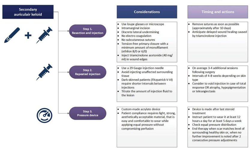
Picture 11. Example Algorithm Governance Keloid.
Prognosis Keloid
Keloid own level recurrence Which tall. Keloid periauricular has a high recurrence rate of around 43%. 22 Recurrence rate keloids are highly dependent on the modality used. Keloid lesions almost never regress spontaneously without therapy. A comparison of recurrence rates of various keloid therapy modalities is shown in Table 2.2.
Table 2.2. Level Recurrence Keloid Based on Therapeutic Modalities. 19
Type | Therapy | Average Level Recurrence | Duration follow up average (month) |
Monotherapy | TAC | 33% | 12 months |
| Excision + radiation | 23% | 14 months |
| Excision + TAC | 15.4% | 12-35 month |
| Excision + therapy compression | 10.6% | 18 months |
| Excision + cryotherapy | 15% | 43 months |
Combination | Excision + Mitomycin C | 16.5% | 6 month |
| Excision + Imiquimoid | 24.7% | 6 month |
| TAC + Laser | 15% | 6 month |
| TAC + 5- FU | 17.5% | 3 month |
| TAC + Cryotherapy | 12% | 6 month |
Prevention Keloid
Until moment This No There is therapy keloid Which own effectiveness 100%. In addition, most keloid cases tend to be recurrent. Thus, education related to keloid prevention is very important in patients who are considered at risk of developing keloids or with a history of keloids. Here are some keloid prevention education that can be given to high-risk patients: 11,19
Patient with People with a previous history of keloids or a family history of keloids should avoid piercing and tattooing.
Patients with acne (especially male patients with acne keloidalis nuchae ) should immediately receive appropriate treatment from medical personnel.
Patients are advised to avoid non-emergency surgery or unnecessary aesthetic procedures.
When a patient has to undergo surgery or other invasive procedures, then the care the wound must be done with Be careful And carefully for avoid formation keloid (use closing wound made from silicone, avoid movements that can make the wound wider, avoid friction on the wound, and keep the wound clean and aseptic).
In addition to the preventive education above, prevention must also be carried out by doctors who may perform surgical or invasive procedures on high-risk patients. keloid. Here are some things what can be done by the surgeon performing the invasive procedure: 11.19
Incision skin must done in accordance tension force skin.
Use instrument Which fine to avoid trauma on skin especially on dark skin areas.
The suture edges should be done by creating minimal skin tension and avoiding excessive use of electrocautery.
Prevent the occurrence infection wound with ensure No There is object foreign left in the wound and give appropriate antibiotics.
Use preventive therapeutic modalities such as compressive, occlusive, injection wound dressings corticosteroids intralesional, or laser For prevent the occurrence of keloids.
Do follow up And take care wound periodically.
REFERENCE
Datema FR, Saridin J, Timmer FCA, Rothuizen LT, Van Zijl FVWJ. Triple Therapy Protocol for Primary and Secondary Auricular Keloids: A Prospective Outcome Evaluation. Dermatologic Surg. 2023;49(9):838–43.
Carvalhaes SM, Petroianu A, Ferreira MAT, de Barros VM, Lopes RV. Assessment of the treatment of earlobe keloid with triamcinolone injections, surgical resection, and local pressure. Rev Col Bras Cir. 2015;42(1):9–13.
Coutinho RLO, Barros BC de, Pedroni EG, Bissoli GP, Kalil M. Giant Keloid of the Ear Lobule: Case Report and Brief Review of Literature. J Dermatology Res Rev Reports. 2022;3(figure 1):1–13.
Choi YJ, Lee YH, Lee HJ, Lee GY, Kim WS. Auricular keloid management in Asian skin: Clinical outcome of intralesional excision and postoperative triamcinolone acetonide intralesional injection. J Cosmet Dermatol. 2020;19(11):3041–7.
Yousef H, Alhajj M, Sharma S, Hospital MF. Anatomy, Skin (Integument), Epidermis. StatPearls. 2023;2–3.
Lopez-ojeda W, Pandey A, Alhajj M, Oakley AM. Anatomy, Skin (Integument) Blood Supply and Lymphatics. StatPearls. 2021.
Faial MMG, Pinnock DD. Earlobe hypertrophy correction. Rev Bras Cir Plástica – Brazilian J Plast Sugery. 2017;32(2):282–6.
Miss L, Shi X. Vascular pathology and hearing disorders. Curr Opin Physiol [Internet]. 2020;18:79–84. Available from: https://doi.org/10.1016/j.cophys.2020.09.004
Nyberg S, Joan Abbott N, Shi X, Steyger PS, Dabdoub A. Delivery of therapeutics to the inner ear: The challenge of the blood-labyrinth barrier. Sci Transl Med. 2019;11(482):1–12.
Zilinsky I, Erdmann D, Weissman Oh, Hammer N, Sora MC, Schenck TL, et al. Reevaluation of the arterial blood supply of the auricle. J Anat. 2017;230(2):315–24.
Betarbet U, Blalock TW. Keloids: A review of etiology, prevention, and treatment. J Clin Aesthet Dermatol. 2020;13(2):33–43.
Limandjaja GC, Niessen FB, Scheper RJ, Gibbs S. The Keloid Disorder: Heterogeneity, Histopathology, Mechanisms and Models. Front Cell Dev Biol. 2020;8(May).
Huang C, Wu Z, Du Y, Ogawa R. The Epidemiology of Keloids. In: Textbook on Scar Management. 2021. p. 30–4.
Liu A.H., Sun XL, Liu DZ, Xu F, Feng SJ, Zhang SY, et al. Epidemiological and clinical features of hypertrophic scar and keloid in Chinese college students: A university-based cross-sectional survey. Heliyon [Internet]. 2023;9(4):e15345. Available from: https://doi.org/10.1016/j.heliyon.2023.e15345
Manoharan A, Rao SM. Analysis of risk factors behind keloids. Int J Res Dermatology. 2020;6(2):138.
Shaheen A. Risk Factors of Keloids: A Mini Review. Austin J Dermatology. 2017;4(2):2–6.
Tsai CH, Ogawa R. Keloid research: current status and future directions. Scars, Burn Heal. 2019;5:205951311986865.
Abdalla MKTM. Applied Basic Science of the Auricular Cartilage. In: Cartilage Repair and Regeneration [Internet]. 2018. p. 203–17. Available from: https:/ /www . intechopen.com/books/advanced-biometric- technologies/liveness-detection-in-biometrics
Elazhary E, Abd Al-Salam F, Abd El-Hafiz H, Maghraby H. Updates on keloids scar pathogenesis, assessment and treatment modalities. J Recent Adv Med. 2022;3(1):75–86.
Ogawa R, Akita S, Akaishi S, Aramaki Hattori N, Dohi T, Hayashi T, et al. Diagnosis and Treatment of Keloids and Hypertrophic Scars—Japan Scar Workshop Consensus Document 2018. Burn Trauma. 2019;7:1–40.
Ojeh N, Bharatha A, Gaur U, Forde AL. Keloids: Current and emerging therapies. Scars, Burn Heal. 2020;6:205951312094049.
Wang JC, Fort CL, Hom DB. Location Propensity for Keloids in the Head and Neck. Facial Plast Surg Aesthetic Med. 2021;23(1):59–64.
The Zorlu Oh, Yazici S, The fight Dear Ş. Keloid formation following ear piercing through the transitional zone. An Bra Dermatol. 2023;98(4):548–50.
Walsh LA, Wu E, Bridges D, Kwan KR, Poondru S, Miller CH, et al. Keloid treatments: an evidence-based systematic review of recent advances. Syst Rev [Internet]. 2023;12(1). Available from: https://doi.org/10.1186/s13643-023-02192-7
Pratiwi IAI, Wardhana M. Right auricular keloid treated with a combination of intralesional excision and corticosteroid injection with tumescent anesthesia: a case report. Medical Science Digest. 2020;11(2):497–503.
Comments