AURICULOPLASTY SECOND STAGE WITH APPROACH NAGATA TECHNIQUE IN MICROTIA CASES
I Made Nudi Arthana
Auriculoplasty stage II for reconstruction with technique Nagata use in microtia cases. Technique Nagata For reconstruction leaf ear stage II indicated when the cartilage is completely revascularized, which is usually about 1 month after the first stage of surgery.
LITERATUR REVIEW
Anatomy ear
Each ear consists of three parts: the outer, middle, and inner ear (Figure 1). The outer and middle ear channel sound waves from the air to the fluid-filled inner ear, amplifying the sound energy in the process. The inner ear contains two sensory systems: the cochlea, which contains receptors for converting sound waves into nerve impulses so we can hear, and the vestibular apparatus, which is important for the sense of balance. 7,8
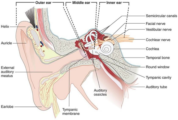
Picture 1. Anatomy Ear 7
Outer Ear
The external ear consists of the auricle (pinna) which captures sound waves. voice, meatus auditory external (channel ear) in where sound waves distributed, And membrane timpani (drum ear) Which move enter and comes out in response to sound. The tympanic membrane is a thin, semitransparent connective tissue, which separates the outer ear from the middle ear. The tympanic membrane is very delicate. The narrow auricle and external acoustic meatus protect it. membrane timpani from injury. As addition Lots follicle hair and gland sweat Which modified (glandula ceruminous) Which located parallel to meatus acoustician external. Follicle the functioning For catch debris And give sensitivity tactile through plexus Which there is on hair roots. Ceruminous gland secretions, called cerumen, slow the growth of microorganisms and reduce the possibility of infection. 9
Middle Ear
Ear middle containing room full air Which called cavity timpani, where the auditory ossicles are located. The tympanic cavity is also connected to the nasopharynx via the Eustachian tube. The pharyngeal tube or Eustachian tube is about 4.0 cm long and penetrates the petrous part of the temporal bone. The connection to the tympanic cavity is narrow and supported by cartilage. The opening to the nasopharynx is wide and funnel-shaped. 9 The Eustachian tube is normally closed, but during swallowing, chewing, and yawning it opens, to maintain equal air pressure on both sides of the tympanic membrane. The three auditory ossicles/middle ear bones: the malleus (hammer), incus (anvil), and stapes (stirrup). The manubrium (handle of the malleus) is attached to the back of the tympanic membrane, its head is attached to the wall of the middle ear, and its processus Which short attached on incus. Appropriate in lower oval window is round window Which Also have layer Which called membrane timpani second. The vibration of the membrane causes the bones to move and transmit sound waves through the tympanic cavity to the foramen ovale. The vibrations then travel through the fluid in the inner ear and stimulate the auditory receptors. The tense part of the membrane is the pars tensa while the slightly tense part is the pars tensa. is pars flaccida. Change atrophy on membrane Because process aging result in membrane more shallow and retraction/stretching. 10
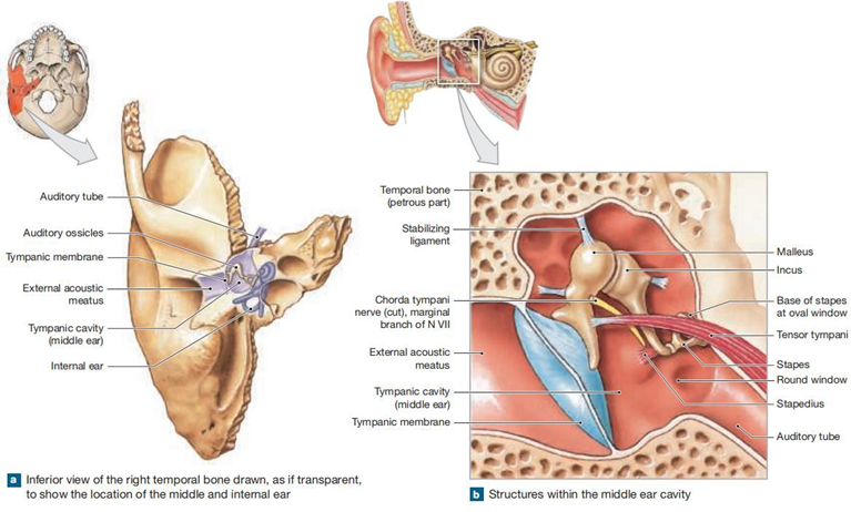
Picture 2. Structure Ear Middle 9
Inner Ear
The inner ear consists of the bony labyrinth, the membranous labyrinth, and the spiral organ (organ of Corti), which is the organ of hearing. The inner ear has a bone-like structure on the outside consisting of the semicircular, vestibular, and cochlear canals, and contains a fluid called perilymph. This fluid surrounds the membranous labyrinth, which is a channel in the bony labyrinth that is the location of hearing and balance receptors. The membranous labyrinth contains a fluid called endolymph which have a level high potassium ions (K+) And level sodium Which low, And on the contrary on perilymph, Which play a role in delivery message. On part middle from ear in there is oval structure Which called vestibule, labyrinth membrane on area This consists of from sacculus and utricle. In the superior and posterior parts of the vestibule are the semicircular canals. The anterior part of the vestibule is the cochlea, a spiral canal that coils almost three times around a bony core called the modiolus, and divides into three channels, namely the cochlear duct (scala media), the scala timpani, And scale vestibule. On membrane basilaris there is organ spiral (organ of corti) Which own cell support And cell hair Which functioning as a receptor hearing. Membrane tectorial is a layer flexible from gelatin that cover cell hair. There is two type cell hair that is cell hair in And Outer hair cells. There are 20,000 outer hair cells and 3500 inner hair cells in each human cochlea. 7
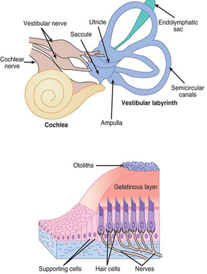
Picture 3. Parts Ear In. 7.9
Ear Embryology
In development ear man, role important located on the branch arch/ branchial arch. Arch This divided by bags Which consists of endoderm on the surface internal and by gap Which consist of ectoderm on its external side. The mesoderm center contains muscles, cartilage, blood vessels, and nerves that will eventually supply and build the surrounding anatomical structures. Molecular signals from the ectoderm result in mesenchymal invasion And is lost gradually from gap And pocket. Importance invasion This mesenchyme is emphasized by the fact that failure of this critical step can result in residual branchial arch And cleft , Which result in various anomaly branchial cleft cysts , for example, represent the persistence of a first branchial cleft . Some authors have also suggested that anomalies in molecular signaling between the ectoderm and mesenchyme may result in failure of chondrogenesis, which may result in varying degrees of anomalies, ranging from the absence of certain components of the auricle to microtia or even anotia in the case of complete failure of chondrogenesis. 4
The development of the human ear begins with the appearance of the otic placenta and vestibulocochlear ganglia at 3 weeks of gestation. The external auditory canal start develop from gap branch First on age 4 Sunday, followed by the formation of a hole by ectodermal proliferation. By 28 weeks, an epithelial core has canalized from medial to lateral, producing a fully patent external auditory canal. Failure of meatus formation external acoustics can causing stenosis or membranous atresia or bones. Development leaf ear started on age pregnancy 5 Sunday with the development of hillocks auricles numbered from 1 to 6, derived from the first (mandible) and second (hyoid) branchial arches. In the sixth week, there are 6 mesenchymal thickenings on the dorsal margins of the first and second branchial arches. Hillocks the first and sixth become more pronounced before the other hillocks , but everything defined on end Sunday 6th. Fusion hillock - those hillocks produce formation leaf ear. Failure fusion hillock - these hillocks can lead to the formation of preauricular or sinus cavities, as well as cleft ears. 4 A thorough understanding of the size and proportions of the “normal” external ear is essential for a reconstructive surgeon. In terms of size, the normal adult ear measures approximately 5.5-6.5 cm, with a width of 50-60% of that size. Along a line connecting the brow and columella, approximately one ear length separates the lateral orbital rim and the root of the helix. 11

Picture 4. Development embryological leaf ear. Migration sixth The auricle hillocks can be seen from (a) the early fetus, through (b) the primitive ear, and finally (c) the fully developed ear. 11
The contribution of each hillock becomes less pronounced in the transition zone between the arches. mandible And hyoid. Although part big agreed that hillock- these hillocks can be followed in development to become specific components of the auricle, some theories doubt this by stating that the hillocks are temporary, representing intensive foci of mesenchymal proliferation, which do not directly lead to the formation of specific components of the auricle. In particular, there is controversy regarding the origin of the development of the ascending helix And crus helical. Along time, contribution each to earlobe has modified, started with His on year 1885. Park proposed that the pinna may develop from hillocks 1 and 6 based on his studies of ear deformities. An additional contribution to the auricular primordium is the free ear fold, which develops behind the second branchial arch and eventually contributes to the helix, scaphoid fossa , and superior crus of the antihelix. Finally, the movements of the auricle in the human embryo were described in detail by Streeter. During development, the external ear gradually move to lateral And dorsal And to direction skull relatively to eyes and mouth. Kagurasho et al determined that ear movement during development is based on changes in the size and shape of the embryo which is also known as growth differential, No on migration from One area to

Picture 5. Origin ear branching 4 .
Table 1. Summary development embryology on ear outside. 4

Abnormalities congenital ear
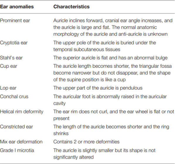
Anomaly ear congenital is results from error during embryological development bone vulnerable auricularis fetus during Sunday fifth until ninth pregnancy or abnormal physical force during development, which affects the normal growth of the ear. The auricle originates from the first and second branchial arches, which give rise to structures such as the auricle, middle ear, inner ear, facial nerve, mandible, maxilla, and hyoid bone. These malformations have been characterized and divided into two separate categories: malformations, which occur early in development (for example microtia, anotia, cryptography) And deformation, which usually occurs in late stages of development and is associated with external compression (eg, lobed ears, protruding earlobes). Because the human earlobe has important aesthetic and cultural value, anomalies in the external ear can significantly affect a child's ability to communicate and socialize. 2
Table 2. Classification Anomaly Ears. 12
Microtia
Microtia is an abnormality of the outer ear (pinna or earlobe and channel hearing external [EAC]). Classification formal has proposed by some expert like Goodbye, Nagata, And Marx. Abnormalities This range from light structure (like ear stand out) until No existence ear outside (anotia). Microtia can occur in isolation or as part of a spectrum of anomalies or syndromes. Although most cases are not associated with a syndrome, the discovery of the underlying gene has helped understand the cause of microtia, especially when it occurs in combination with other defects of the head and face. 2,3
Microtia is condition in where leaf ear No develop fully, covering a wide range of congenital anomalies that can vary from mild to non-significant. existence ear same outside once (anotia). Microtia more general occurs in men, with ratio man And Woman around 2.5 appeal 1. Approximately 77-93% of cases involve involvement of only one ear, with approximately 60% case influence ear right. Microtia bilateral, Which involving both ears, found in about 10% of patients. More than two-thirds of cases of severe microtia or anotia are usually associated with congenital aural atresia (CAA), in which the external ear canal is closed. However, although the ear deformity may appear minor, this does not exclude the possibility of problems with the middle ear, inner ear, or facial nerve. 1,2

Picture 6. Spectrum presentation microtia from ear small to anotia. (a) Marx grade 1 or Nagata Microtia atypical type. (b) Nagata Microtia concha type or Marx grade 2 (c) Microtia concha type Marx grade 2 or Nagata concha type microtia small (d) Marx grade 3 or Nagata microtia type lobule (e) Marx grade 3, or Nagata's anotia. 11
HEAR MAPS is method summary For classify aspect ear and face in patients with microtia. This is the only system that includes hearing function. bone conduction and air conduction hearing levels. Inspection This Also evaluate level severity remainder ear, score atresia which is determined by CT scan, size remainder lobular, deviation mandible, level soft tissue hypoplasia and facial nerve paralysis. This examination also identifies the presence or absence of associated syndromes. HEAR defines the microtic ear, while the MAPS from classification referring to on face. HEAR represent hearing, ear class, atresia score, and residual lobes. MAPS refers to the face with letters representing mandible, soft tissue asymmetry, paralysis, and syndrome. This system provides the most concise and comprehensive phenotypic description of individuals with microtia. The term "constricted ear " was coined by Tanzer to categorize a group of congenital ear deformities. Tanzer noted the subtle finding of a hood helix And alignment antihelix And enter lop ear And cup ear to in the scoring system for constricted ears. It is debatable whether some ear Which narrow is microtia level 1 Which Actually versus category abnormality form ear Which separated. Description original Tanker covers three groups and two subgroups. Group 1 constricted ears only close at the helix or lop portion of the ear. Group 2 involves the helix and scaphus. This form of constriction involves the hood and gives a "cup" appearance. ears), And shared Again become two subgroup. Group 2A Enough moderate and does not require additional skin to expand the helix. 11
Table 3. HEAR MAPS is method summary For classify aspects of the ear and face in patients with microtia. This is the only system that includes a listener function

Picture 7. System HEAR classification MAPS developed For evaluate microtia and consider abnormality And syndrome craniofacial Which related. (a) This patient is classified as H1.6E9A1R1 M2A1P1S1. Conduction normal bone in the range of 10-20 dB with air conduction in the range of 60-70 dB. He had grade 3 microtia with a Jahrsdoerfer atresia score of 9 with CT scan. He doesn't have any craniofacial anomaly other related and there is no evidence of a syndrome. (b) This patient is classified as H1.6E3A3R1 M2A3P1S1. He has microtia grade 3 with Jahrsdoerfer atresia score 3 on CT scan. His earlobe is the same size as his right ear. The mandible is slightly displaced with significant soft tissue hypoplasia. He has no facial nerve deficits and no evidence of any syndrome. (c) This patient is classified as H1.6E3A4R1 M1A1P4S1, He has Score atresia The Driver 4, paralysis mandible marginal And there is no evidence of the syndrome. 11
Table 4. Classification Tanker.11

Reconstruction auricle (auriculoplasty) stage II with technique Nagata.
Technique Nagata For reconstruction leaf ear stage II indicated when the cartilage is fully revascularized, which is usually about 1 month after the first stage of surgery. This technique involves implanting cartilage in the postauricular area and enlarging the contact area of the severed cartilage with the muscle base using an inversion maneuver. This improves nutrition and maintains the shape and function of the auricle. The skin is removed but the perichondrium is preserved to reduce cartilage curvature and flattening. Incision For lift skin mastoid own long around 4 cm, minimizing damage to surrounding tissue and allowing secondary reconstruction if necessary. 13
In 1993, Nagata published a modified two-stage approach. For microtia autologous in where lobule transposed on the stage that The same with fabrication framework (including construction tragus) And placement. With thus, Which First from Nagata First combines the first, second, and final stages of Brent's technique. In addition, both authors proposed their own classification systems to describe the variable appearance of microtia deformities. While Brent acknowledged the possibility of simultaneous lobular transposition and skeletal placement, and demonstrated results tragus Which more Good with technique This, He state that transposition lobule more Good reserved as procedure secondary. Good Nagata and Firmin (initially) do transposition lobule And use posterior surface of the lobe to line the tragal support of the skeleton in the first stage. Indeed, construction of the tragus and inter-tragal notch is a fundamental improvement Which put forward by Nagata. In technique Which presented by Tanzer, an incision was used, which extends from the posterior surface of the lobule to the mastoid surface in a V shape, and the lobule is displaced posteriorly. The modification of the incision line presented by Nagata evolved from the original incision. shaped V become incision big shaped W, so that create four skin flaps. Points A and B, located 1 cm from the center of the W-shaped incision, are sutured together to create a cone of skin to line the recess of the inter-tragal notch. The center of the skin flap formed by the W-shaped skin incision serves as a subcutaneous pedicle. Nagata recommends preserving the subcutaneous pedicle to increase vascularization of the distal part of the mastoid flap, which will cover the concha and the posterior surface of the tragus. On the anterior surface of the lobule, Tanzer uses a straight-line incision for transposition lobule. Nagata modify line incision For to form flap lobular skin anterior, skin front And flap tragus. With thus, formed anterior and posterior lobular skin flaps, an anterior tragus flap, and a mastoid skin flap. Incision circular small (2 mm) flap transposition lobule is basis for the formation of the attachment point for the inter-tragal notch . Tanzer uses a V-shaped incision to achieve lobule transposition. In this process, the surface skin posterior lobule And mastoid Which close together No utilized, and usage grafting skin conch become a need. Nagata make a fundamental contribution to skin sheath management by increasing the surface area available skin surface to cover the cartilage construct. This is accomplished by lowering a W-shaped incision line and using a posterior skin flap to encompass the anterior portion of the framework, thereby eliminating the need for a skin graft in the first stage. For conchal microtia, the same posterior incision is used as for lobular microtia. However, a modification of the incision is made along the anterior residual surface. The incision line for the anterior surface extends from the helix posterior to the lesser conchal notch, and the terminal portion of the incision is removed circumferentially to form a U-shaped inter-tragus notch. 11
Brent take notes that side negative from technique Nagata is that The lobule appears less natural because most of the posterior lobe is used to cover the concha and the posterior aspect of the tragus. The Nagata technique has been adopted as the leading technique for autologous microtia reconstruction. However, a thorough understanding of the three-dimensional architecture of the normal ear is required to reproduce adequate results. Nagata uses the sixth to ninth costal cartilage segments for frame fabrication, leaving the posterior perichondrium in situ. The basic frame block is made from the seventh cartilage And eighth bone vulnerable costa, temporary part Which remaining used for to form anti-helix, crura inferior And superior, tragus, And curve inter-tragus. Nagata generates an anti-helical complex from additional rib cartilage and mounts it to the basic skeletal block, resulting in better definition of the anti-helix than Brent's technique. The helical and crus helicis units are constructed from the ninth costal cartilage, with the crus helicis extended to the posterior surface anti-helix For define Cymba And cavity conchae. Reconstruction was performed after the age of 10 years, with the formation of a skin sheath adapted to Nagata's skeleton. Nagata emphasized the conservation of the entire perichondrium. as step integral in preventing deformities chest wall And reach regeneration bone vulnerable, allow reconstruction secondary or in cases of bilateral microtia. 11
The Nagata technique is one of the leading techniques in microtia reconstruction. in a way autologous. However, technique This need understanding Which details about the three-dimensional architecture of the normal ear. After thoroughly understanding the morphological features of the auricle, the reconstruction results can resemble the normal ear. The main contribution to the creation of the framework involves features such as the inter-tragal notch, tragus, and crus helicis associated with the cymba and cavum conchae. For make framework, bone vulnerable from segment bone costal fragility sixth until ninth harvested, leave perichondrium posterior in its place. Part base framework made from bone vulnerable seventh And eighth, while the other parts of this costal cartilage segment are used to form the anti-helix and the inferior and superior crura, as well as the tragus and inter-tragus notch. Nagata use addition bone vulnerable ribs For form a complex anti-helix And install it to block base framework, than carving the anti-helix into the underlying framework as in the Brent technique. This results in better definition of the anti-helix. The framework units are held together with gauge wires at specific intervals. The sixth costal cartilage segment is saved for use during the second stage of reconstruction. 11
In the second stage, the formation of the retro-auricular sulcus is performed. Healing is carried out by making incisions along the edges of the construction, Then put flap temporoparietal fascia and skin grafts . Flap The fascia is then passed through the subcutaneous tunnel to cover the posterior surface of the reconstructed ear. This technique has been shown to be successful in producing cartilage regeneration, providing a suitable material for secondary reconstruction or in cases of bilateral microtia. The second stage in the microtia reconstruction process is the formation of the retro-auricular sulcus, which is usually performed approximately 6 months after the fabrication and placement of the framework. An incision is made along the periphery of the construct, approximately 5 mm from the edge of the auricular helix. Initially, Nagata used a technique of grafting skin to the posterior surface of the framework, similar to the method used by Brent. However, he later added the use of semilunar costal cartilage blocks and concave costal cartilage which is attached with nylon sutures 4-0 to surface posterior framework, as high as wall concha posterior. Part last of process This closed with flap fascia temporoparietal And graft skin. Flap fascia taken through incision zigzag in skin For avoid damage on hair follicles. Then, a fascial flap is placed through the subcutaneous tunnel to the posterior surface of the reconstructed ear. The retro-auricular skin is then pulled toward the sulcus, and a full-thickness skin graft is used to cover the remaining surface. Nagata later modified his technique by combining return flap fascia temporoparietal with graft skin Which taken from the ipsilateral scalp in split thickness. 11

Picture 8. Auriculoplasty stage II Nagata 11
Chen and colleagues describe a modification to Nagata's second stage, in which graft skin with thickness separated lifted in continuity with full-thickness skin over the anterior surface of the reconstructed ear for cover area retro-auricular And create coverage skin Which continuous on the ear. Nagata's technique for the second stage of autologous auricular reconstruction represents many important improvements over Tanzer and Brent's approach to formation sulcus retro-auricular. Brent propose elevation ear And with split-thickness skin grafts . However, significant effacement of the sulcus and loss of auricular projection resulted. Nagata advanced the concept of placing cartilage graft segments along the surface posterior anti-helix For add wall posterior conch thus contributing to ear projection and preventing retraction of the retro- auricular sulcus. 13
Wang et al. explained that the combination of network expansion and Nagata technique provides approach Which effective And efficient For reconstruction auricular in patients microtia. Strategy two stage allow expansion flap in a way gradually, producing an appropriate area and thickness for the reconstructed ear. This technique produces satisfactory medium-term results, with patients and their families expressing satisfaction with the clear lines, color, and in accordance, projection Which Good, And symmetry ear Which reconstructed. This procedure Also allow reconstruction simultaneous leaf ear And tragus. However, there is potential complications Which related with technique This, including necrosis ischemic on the distal earlobe and skin of the conchal cavity, hematoma, and the need for further restoration in some cases. In the first stage, an expander is inserted to area mastoid And in a way gradually expanded during period a certain amount of time. This allows the flap to reach the appropriate area and thickness for reconstruction. After a resting period of almost 2 months, the expander is removed and the framework bone vulnerable three layer entered to in pocket through incision lobules shaped Y on stage second . Reconstruction simultaneous leaf ear And tragus also performed during the second stage. This two-stage strategy combines tissue expansion with the Nagata technique, resulting in an effective and efficient approach to auricular reconstruction. 14
Wu et al. reported a series of nine microtia reconstructions in one stage use bone ribs with implant For to form auricular skeleton composite or hybrid. They to form bone vulnerable ribs the ninth into a helix and sutured it to an auricular implant prosthesis (Stryker, Kalamazoo, MI, USA), and reported acceptable cosmetic results without any extrusion during monitoring during 2 year. Furthermore, Money et et al., Kim et et al., And Shan et a. use implant (Stryker) as buffer behind the auricle to support the auricular framework of the ribs, and cover the behind the auricular defect with vascular temporoparietal fascia using classical reconstructive techniques. Nagata. Their modifications avoid taking bone ribs addition during stage second reconstruction And achieve projection auricular Which can accepted without extrusion implant furthermore. This report shows that the use of implants combined with ribs minimizes the risk of extrusion. In our study, we hypothesized that there is no difference in the esthetic results of Nagata microtia reconstruction using ribs compared to the hybrid approach, both have comparable complications, but the hybrid approach requires fewer ribs. 15,16

Picture 9. Technique surgery For reconstruction framework auricularis with rib cartilage alone or a combination of rib cartilage and porous polyethylene. 17
In the literature search, there were no studies comparing QoL between patients who received autologous and alloplastic reconstruction. Some have studied QoL after ear reconstruction using a specific technique, such as those conducted by Widodo et al., who found that many patients had good health-related quality of life (HRQoL) after ear reconstruction using the Nagata technique (autologous), and Braun et al., who found that alloplastic reconstruction using MedPor® significantly improved patients' HRQoL. These findings are supported by a systematic review stating that both autologous and alloplastic reconstruction have good outcomes in HRQoL. However, studies comparing QoL after autologous and alloplastic reconstruction have not been conducted, so no conclusions about the superiority of one technique can be drawn at this time. This suggests the need for further research to study how the two techniques may affect patients' lives differently. Analyses comparing preoperative and postoperative QoL, as well as autologous and alloplastic reconstruction, would be very beneficial. 5
REFERENCE
Bartel-Friedrich S. Congenital Auricular Malformations: Description of Anomalies and Syndromes. Facial Plast Surg. 2015;31(6):567–80.
Liaw J, Patel VA, Car MM. Congenital anomalies of the external ears. Opera Tech Otolaryngol - Head Neck Heaven [Internet]. 2017;28(2):72–6. Available from: http://dx.doi.org/10.1016/j.otot.2017.03.012
Joukhadar N, McKee D, Caouette-Laberge L, Bezuhly M. Management of Congenital Auricular Anomalies. Plast Reconstr Surg. 2020;146(2):205e- 216e.
Moneta LB, Quintanilla-Dieck L. Embryology and anatomy of the ear. Oper Tech Otolaryngol - Head Neck Surg [Internet]. 2017;28(2):66–71. Available from: http://dx.doi.org/10.1016/j.otot.2017.03.011
Widodo DW, Irfan I, Safitri ED, Reksodiputro MH. Outcomes of Autologous Reconstruction in Comparison with Alloplastic in Microtia Patients. Indonesian Medical eJournal. 2023;10(3):258–64.
Yamada A. Autologous Rib Microtia Construction: Nagata Technique. Facial Plast Surg Clin North Am. 2018;26(1):41–55.
Barrett TO, Barman SM, Brooks HL, Yuan J. Hypothalamic Regulation of Hormonal Functions. Vol. 148, Ganong's Medical Physiology. 2019. 698–
Hall JE. Guyton and Hall textbook of medical physiology / John E. Hall.— Thirteenth edition. p. UNITS. Guyton and Hall textbook of medical physiology / John E. Hall.—Thirteenth edition. p. UNITS. 2016. 5–24 p.
Martini, Frederic Tallitsch RB, Nath, Lindsley J. Contents in Brief. Vol. 27, Journal of Vascular and Interventional Radiology. 2018. A4 p.
dee-unglaub-silverthorn-human-physiology-an-integrated-2019.
Reinisch JF, Tahiri Y. Modern microtia reconstruction: Art, science, and new clinical techniques. Modern Microtia Reconstruction: Art, Science, and New Clinical Techniques. 2019. 1–342 p.
Chen Y, Wang W, Wang Y, Mao X. Using Ear Molding to Treat Congenital Auricular Deformities. Front Pediatr. 2021;9(December):1–6.
Ly Y, Mom L, To C, Zhang W, Liu M. A two-stage inversion technique for total auricular reconstruction: Case report and literature review. BMC Surg. 2018;18(1):10–4.
Wang B, Guo R, Li Q, Ou Y, Hu J, Wang Y, et al. A novel two-stage strategy combing tissue expansion and Nagata's technique for total auricular reconstruction. J Plast Reconstr Aesthetic Surg [Internet]. 2021;74(9):2358–63. Available from:731 p. https://doi.org/10.1016/j.bjps.2020.12.087
Kim YS. The use of medpor as a projection block for the elevation of the constructed auricle in total auricular reconstruction. JPRAS Open [Internet]. 2017;13:53–61. Available from: http://dx.doi.org/10.1016/j.jpra.2017.05.011
Shan J, Guo Y, Chang KW, Zhang T. A modified technique for firm elevation of the reconstructed auricle. Eur Arch Oto-Rhino-Laryngology. 2016;273(10):3019–24.
Ku PKM, Vlantis AC, Tong MC, Chan TTT, Yeung ZWC, Cho RHW, et al. A Hybrid Auricular Framework of Autologous Rib Cartilage and a Porous Polyethylene Implant for Reconstruction of Congenital Microtia: A Modification of Nagata's Technique. Facial Plast Surg Aesthetic Med. 2024;26(1):15–22.
Jhawar M, Yadav S, Prasad S, Tiwari D, Patni D, Munjal VR. A case of microtia with bilateral conductive hearing loss with unusual findings. Int J Otorhinolaryngol Head Neck Surg. 2023;9(6):501–4.
Alkhamisy A, Gb A, Alyousef G, Alseddiki FA, Morjan M. Bilateral congenital chest wall defects associated with microtia: First case report. J Pediatr Surg Case Reports [Internet]. 2022;86(August):102438. Available from: https://doi.org/10.1016/j.epsc.2022.102438
Chwa ICE, Laub PJ, Yamada A. Demystifying Small Concha type Microtia: Advancements in Classification and Surgical Approach. Plast Reconstr Surg - Glob Open. 2022;10(11):1–4.
Bulstrode N, Stewart K, Yamada A. Celebrating the life of Satoro Nagata, MD, a microtia pioneer, 1950-2022. J Plast Reconstr Aesthetic Surg [Internet]. 2022;75(2):509–10. Available from: https://doi.org/10.1016/j.bjps.2022.02.010
Mulcare D. Prosthetic rehabilitation of unilateral congenital microtia with implant-retained auricular prosthesis - a case report. J Ir Dent Assoc. 2022;68(1, February/March):39–43.
A Two-Flap Combination for Auricular Elevation in Microtia Reconstruction. Plast Reconstr Surg (Plastic Reconstr Surgery). 2023;151(6):pp 991e-1001e.
Comments