VOCAL FOLD PARALYSIS
I Made Nudi Arthana
Vocal fold paralysis (VFP) has been defined as the absence of movement or paralysis, resulting in loss of neural input. Electrophysiological investigations have shown that VFP is actually a heterogeneous clinical condition and is perhaps best thought of as a continuum of neurogenic dysfunction, encompassing partial denervation, complete denervation, variable degrees and patterns of reinnervation. It should not be surprising that paresis or incomplete paralysis in which some gross vocal fold mobility is retained has been considered alongside paralysis as a clinical entity 1
LITERATURE REVIEW
Anatomy Larynx
Structure Buffer Larynx
Larynx is a structure shaped tube Which formed from a system Which complex Which consists of from muscle, cartilage, network tie. Larynx hang from bone hyoid, Which is the only one bone in in the body that does not articulate with other bones. The skeleton of the larynx is composed of on 3 cartilage Which in pairs And 3 cartilage Which No paired. Cartilage thyroid is cartilage No in pairs Which the biggest, located under the hyoid bone, and hanging on ligamentum. The thyrohyoidum is two alae or wings of the thyroid cartilage that are shaped like shields. The most anterior part of this cartilage is often prominent in some men, and is usually called as the “ Adam's apple” . At the posterior edge of each alae , there is a superior cornu And inferior. Articulation corn inferior with cartilage cricoid, allow A little shift or movement between cartilage thyroid And cricoid 4. The second unpaired cartilage is the cricoid cartilage, which is also easy palpable in lower skin, attached on cartilage thyroid past cricothyroid ligament, its shape often depicted as A “ signet ring" which is shaped circle full And not capable expand. Intubation endotrachea the old one often damage the mucous layer of the ring and can causing subglottic stenosis obtained. Cartilage third Which No in pairs is epiglottic cartilage, Which shaped like A bat ping pong. Grip or The petioles are attached via a dependent ligament to the thyroid cartilage just above the vocal cords, temporary part raccoon expand to on in behind corpus hyoideus into the lumen pharynx, separate base tongue And larynx. Adhesion from The epiglottis allows cartilage the For invert, A movement Which can form For push food And fluid in a way direct to in the esophagus And protect cord vocalist And road breathing during process
swallow 4 .
The three paired cartilages are the arytenoid, cuneiform, and corniculate. Arytenoid shaped like pyramid And Because they attached on the vocal cords, allowing the opening and closing movements of the vocal cords which are important for respiration and vocalization. The cuneiforms and corniculates are very small and have no apparent function (J. Dance Jr, Milton, 2011). Figures 1 and 2 below illustrate the structures of the larynx from the lateral and medial sides 5 .

Picture 1. Structure larynx 5
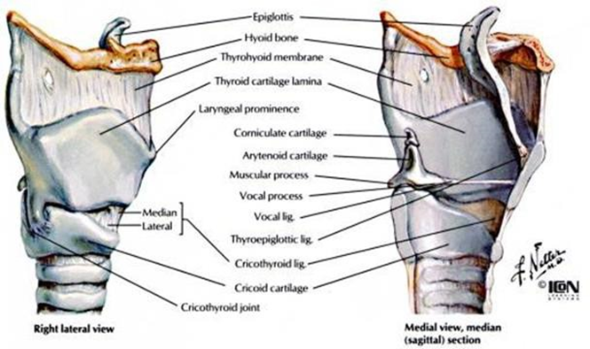
Picture 2. Structure Larynx Side Lateral And Media 5
Muscle
The muscles of the larynx consist of two main groups, namely the extrinsic muscles and the intrinsic muscles. The extrinsic muscles of the larynx are the muscles of the laryngohyoid complex which function to raise, lower, or stabilize larynx. It is called muscle extrinsic Because muscle This in One attached to the larynx and also attached outside the larynx. While the intrinsic muscles are muscles that are anatomically limited to muscles that are attached directly to the larynx. The intrinsic muscles modify the size of the opening in the glottis along with the length and tension of the vocal folds 4 .
Muscle Extrinsic
Muscle extrinsic attached on surface larynx And surface outside larynx. Extrinsic muscles function to move the larynx. Because Os.hyoideum is connected with larynx by membrane hyothyroidism And by epiglottis, then the muscles that move the Os.hyoideum will also move the larynx. There are 8 extrinsic muscles of the larynx, divided into 5 :
Muscle Suprahyoid
Functions to lift the larynx upwards. Consisting of M.Stylohioid, M.Myohioid, M.Geniohioid, and M.Digastric.
Muscle Infrahyoid
Functions to pull the larynx downwards, consisting of M.Sternothyroid, M.Sternohioid, M. Thyrohyoid, And M.Omohyoid. Muscles This play a role on the movement and fixation of the larynx as a whole. Consists of the elevator and depressor muscle groups. The depressor muscle group consists of the muscles, thyrohyoid, sternohyoid, and omohyoid which are innervated by the ansa hypoglossus from C2 And C3. Group muscle elevator consists of from The anterior and posterior digastric muscles, stylohyoid muscles, geniohyoid muscles and myohyoid muscles are innervated by cranial nerves V, VII and IX. This group is important in the function of swallowing and phonation by lifting the larynx below the base of the tongue.
Muscle Intrinsic
Contraction of intrinsic muscles is related to the movement of the vocal cords. The intrinsic muscles of the larynx function to maintain and control the respiratory airway through the larynx, control resistance to expiratory air during phonation and assist the function sphincter in prevent aspiration foreign object during process swallow. The intrinsic muscles consist of the posterior cricoarytenoid muscle, lateral interarytenoid muscle, cricothyroid muscle and thyroarytenoid muscle .

Picture 3. Muscles Larynx 5
Picture 3 explain about muscles larynx, in where M.Cricothyroid located on the front surface of the larynx, namely: between the sides lateral cricoid and thyroid cartilages. This muscle functions to narrow the cricothyroid space anteriorly and this movement enlarges distance between cartilage thyroid And cartilage arytenoid, Which hitchhiking on the cricoid. Adhesion anterior and posterior vocal ligaments separated further away. The end result is lengthening and stretching of the vocal cords. Contraction of the posterior cricoarytenoid muscle brings the muscular processes of the arytenoids backward and to rotate process vocalist to lateral. Muscle This functioning as abductor main vocal cords. The lateral cricoarytenoid muscle performs adduction of the vocal cords. The external thyroarytenoid muscle works to adduct the vocal cords, and also changes the tension And thickness edge free voice. Sphincter glottis interesting cartilage arytenoid to front For reduce tension ligament vocalist and enlarge tape thickness voice. Muscle This innervated in a way bilateral by nerve laryngeal recurrent, therefore there is no paralysis due to disease affecting the unilateral recurrent nerve. This muscle also receives motor innervation from the superior laryngeal nerve. The aryepiglottis muscle functions to cover the superior laryngeal sphincter, but its shape small and often almost none. Muscle This can become hypertrophied if the function of the false vocal cords replaces the function of the real vocal cords 4 .
Nerves, Bleeding And Drainage Lymphatic Larynx
There are two pairs of nerves that innervate the larynx with sensory and motor innervation, namely: two superior laryngeal nerves and two inferior or recurrent laryngeal nerves. The laryngeal nerves are branches of the vagus nerve. The superior laryngeal nerve leaves the vagus trunk just below the ganglion nodosum, curves anteriorly and medially below the external and internal carotid arteries, and bifurcates into an internal sensory branch and an external motor branch. The internal branch penetrates thyrohyoid membrane to take care of the innervation sensory vallecula, epiglottis, sine piriformis, And all over mucosa internal superior larynx free edge of the true vocal cords. Each external branch is supply motor For One muscle just, that is muscle cricothyroid. In Inferiorly, the recurrent nerve passes up in a groove between the trachea and esophagus, entering the larynx proper behind cricothyroid articulation, and innervates the motor nerves of all the intrinsic muscles of the larynx except the cricothyroid. The recurrent nerve Also look after sensation network in lower cord vocalist true (region subglottis) and superior trachea. Neurological journey Different right and left recurrences also show different pathways higher neural than the laryngeal innervation. Because journey nerve recurrence left Which more long as well as the relationship with the aorta, this nerve is more susceptible to injury than the right nerve 4 .
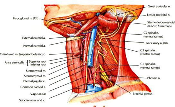
Picture 4. Nerves And Bleeding On Larynx 4
Figure 4 explains the innervation and blood supply of the larynx. artery And drainage venous from larynx parallel with supply his nerves. The superior laryngeal artery and vein are branches of the superior thyroid artery and vein, and they join the internal branch of the superior laryngeal nerve to form the superior neurovascular pedicle. The inferior laryngeal artery and vein arise from the inferior thyroid vessels and enter the larynx with the recurrent laryngeal nerve 6 .
There are two separate drainage systems, superior and inferior, with the dividing line being the true vocal cords. The vocal cords themselves have a poor lymphatic supply. Superiorly, the lymph flows along the superior neurovascular pedicle to join the superior lymph nodes of the deep cervical chain at the level of the hyoid bone. The subglottic drainage is more diverse, including the pretracheal nodes (one node is located just anterior to the cricoid and is called the Delphian node), the inferior deep cervical nodes, the supracalvicular nodes, and even the superior mediastinal nodes 6 .
Physiology Larynx
The larynx is the organ that produces sound, and has other main functions for airway protection, respiration and phonation. Voice is the sound produced when lung air is exhaled through the vocal cords that are somewhat close together. Air forces the separation of the true vocal cords. Because it will reduce the subglottic pressure, the vocal cords will bounce to close together again. Repetition fast, 125 time on man And 250 time on woman will causes vibrations of the pharyngeal air, which produces the sound of the human voice 4 . The basic pitch of a voice is determined by the length and tension of the vocal cords. The pitch varies in accordance frequency its vibrations. The Hardness voice depends on pressure which is formed under the vocal cords. The sounds emitted by the larynx form vowels. The different vowels are determined by the way the pharynx and oral cavity shape them to resonate the sound 7 .
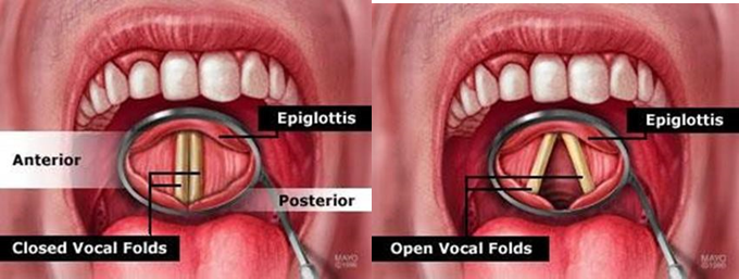
Picture 5. Condition tape voice moment open And closed 8
Other alternative mechanisms exist to form a variable air column in the pharynx. In some cases, for example, patients can speak by bringing their false vocal cords close together to vibrate. After laryngectomy, patients can speak by swallowing air into the esophagus and causing it to vibrate with the pharyngoesophageal tissue 9 .
Sound is converted into speech by stopping the air flow to to form consonant. Production saying Which can understood depends on coordination neuromuscular between cortex motor And cerebellum as well as system muscle pharynx, palate, tongue and lips. These tools are structures that stop the flow of air 10 .
Singing requires integrated production of pitch and volume at the glottis. harmonious, Which relate with mechanics mouth And pharynx, and in accordance with the desired rhythm. The quality of sound in the voice, speaking, and especially singing depends on the additional tones formed in the larynx. This is a mathematical multiplication of the fundamental frequency of the vibrating structure. Vibration tape voice nature complex And combination various vibration as well as various additional tones 10 .
Paralysis Plika Vocalist (PPV)
Definition Paralysis Plika Vocalist (PPV)
Motor innervation of the larynx can cause damage to the neck and mediastinum. Area Which damaged usually in the area arch aorta (in side left) or subclavian artery (on the right side), or in the tracheoesophageal groove. Paralysis means the impairment of the ability of a limb to move and function, usually due to nerve damage. Paralysis can also occur in the vocal cords. PPV happen consequence the disappearance motion active from tape voice And can happen on one or both vocal cords which cannot open or close properly should. PPV defined as lost adduction/abduction normal due to lesions distant from the vocal cords, as a result of nerve injury to the recurrent laryngeal nerve (RLN) 3 .
PPV has many known causes, including trauma, malignancy, and surgery. PPV Can nature idiopathic but Can Also serve description of many underlying serious disease processes. Mechanical fixation of the cricoarytenoid joint can happen after dislocation cricoarytenoid, inflammation local, or invasion neoplastic. Term PPV used For show disturbance laryngeal movement in a way total or part Because injury neurological, Which can affecting the larynx unilaterally or bilaterally. Several studies have shown that the frequency of various causes of PPV changeable. Etiology of paralysis the larynx has changed from time to time, with decline prevalence malignancy And improvement recently This on case Which due to by operation thyroid 3 .

Figure 6. Plika Position Vocalist 3
Figure 6 illustrates the position of the vocal folds, where the vocal folds are considered adducted if the position of the vocal folds is along the median line (c) or paramedian line (d), and are said to be neutral if the position of the vocal folds is along the intermedian line. (b), And abduction when plika vocalist on line lateral (a) abduction partial or (e) full abduction.
Etiology Paralysis Plika Vocalist (PPV)
Vocal cord paralysis can be caused by various diseases or surgery. There are many studies on PPV but the exact incidence of PPV is still difficult to determine. A study conducted by Wang, et al., showed that the incidence of PPV increases with increasing patient age. The peak incidence is between 51 and 70 years. This may be a result of increased incidence of tumors and damage neurological on individuals Which aging. More male patients than females have unilateral or bilateral PPV, possibly because of an increase in postoperative cases resulting from surgery for lung and throat cancer. on man. PPV left happen around two time more often than Right PPV. These results are comparable to previous studies. Studies have shown that neoplasm, surgery, and idiopathic paralysis are the 3 main causes of UVFP 11 .
Study Which done by Wang, et al. state that one hundred Ninety-four patients with PPV were identified, including 178 cases with unilateral PPV. And 16 with VFPB. Age average moment diagnosis is 61 year with a range from 16 to 94 years. For patients with UVFP, left PPV was diagnosed in 122 (69%) patients and right vocal cord paralysis in 56 (31%) patients. One hundred thirteen (58%) patients were male and 81 (42%) were female. Surgery is reason most general from PPV, counted approximately 119 patients (61.3%) patients. Surgical procedures included thyroid surgery for 56 patients, throat surgery for 44 patients, thoracic surgery (lung and heart surgery) for 13 patients, and spine surgery for 3 patients 3 .
Pathophysiology Paralysis Plika Vocalist (PPV)
In the larynx area, anatomically there is the vagus nerve and its branches, that is nerve laryngeus superior And nerve laryngeus inferior or recurrent that innervates the vocal cords. If there is pressure or damage to this nerve, paralysis of the vocal folds occurs, where the vocal cords cannot adduct. Normally, when phonating, both vocal cords adduct, but because of paralysis of one or both vocal cords, the vibrations produced by the vocal cords are not optimal 4 .
In general, there are five positions of the vocal cords according to the degree of the laryngeal ostium: median, paramedian, intermedian, slightly abducted, and fully adducted. If the paralysis occurs bilaterally, this position is indicated by observing the size of the glottic gap. If the paralysis occurs unilaterally, the observation must first estimate the actual midline position and then relate it to the position of the vocal cords. Each lesion along the course of the recurrent laryngeal nerve can cause laryngeal paralysis. Lesions intracranial usually accompanied by symptoms other And more manifest as disturbance neurological And No disturbance voice or articulation. Brainstem lesions primarily cause voice disorders, but may be accompanied by other neurological signs. The position of the vocal cords is the single most important factor, and the clinical symptoms of paralysis vary depending on on position tape voice, that is: median, paramedian, intermediate, abduction A little, and abduction full 4
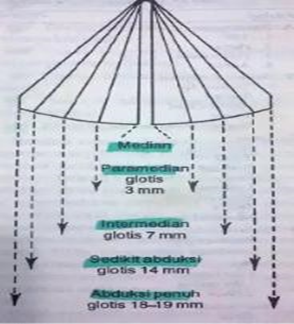
Picture 7. Position Tape Voice 4
Unilateral paralysis in the median position, found in long-standing recurrent nerve paralysis. On examination, the paralyzed vocal cord appears somewhat atrophic and is positioned slightly lower than the normal vocal cord, but on phonation it appears nearly normal. Arytenoids on side Which paralysed leaning to front. The symptoms usually unclear And voice normal on talks. Voice Which need Wide changes in pitch, such as when singing, will be disrupted. During heavy physical exercise, there will be shortness of breath and stridor 4 .
Unilateral paralysis in the paramedian position is a common result of new recurrent nerve palsy. The degree of dysfunction is greatly influenced by the degree of compensation achieved. Examination of the larynx shows paramedian vocal cord paralysis. The membranous vocal cord is usually slightly curved and lower than the normal vocal cord. The paralyzed vocal cord appears to bulge upward on phonation and the glottis remains somewhat oval in shape. The arytenoids appear to cross the midline and move behind or in front of arytenoids paralysis, if the paralysis has been going on for several days. Symptoms in cases that do not experience compensation for paramedian paralysis include: voice hiss, hoarse, time phonation shorten, volume voice, And reduced pitch level, as well as diplophonia. If compensation occurs, the symptoms will reduce And voice will become normal return on a number of cases. There is usually a slight dysphonia and in some cases an abnormally high pitch "falsetto", due to compensatory efforts for the elongated glottis. Usually in older people there is no compensation for this vocal cord position 4 .
Paralysis bilateral on position paramedian is consequence which is commonly found in recent bilateral recurrent nerve paralysis. The symptoms very varies on each individual And in the form of dyspnea and stridor. Dysphonia is inversely related to dyspnea and stridor. Dysphonia is characterized by a weak, slightly hoarse, breathy voice accompanied by volume disturbances. voice And change tone. Should, dyspnea No clear on rest time, but physical work usually causes a little inspiratory stridor and difficulty breathing. By examining the larynx this condition can be revealed. Usually wide glottis in commissure posterior 3-4 mm. Tape voice usually slightly curved again, and on expiration the superior part bulges 4 .
Paralysis bilateral on position median, can happen quick after injury to the recurrent laryngeal nerve, or may be delayed up to 20 year. Symptom Which clear is dyspnea And existence stridor inspiration. Patients tend to For reduce his activities And still silent For get enough oxygen for their needs. An upper respiratory tract infection can cause blockage larynx total, like Also on a the stimulus that cause inspiration in with suddenly. Blockage suddenly on inspiration is caused by adduction of the vocal cords, due to the aerodynamic effect of the airflow striking the superior surface of the vocal cords and pushing them medially. Because of this danger, patients usually breathe shallowly and slowly, and avoid physical exertion or stimulation. The voice remains Good, And most patient deny that There is change voice. However, fine vocal functions, such as singing, are impaired. When examined when phonation, larynx it seems normal, but tape voice No can abducted from position in line middle on time inspiration, so that the airway is only a thin oval-shaped gap. In some cases, the airway is subjectively adequate, due to the difference in the height of the vocal cords 4 .
Paralysis tape voice on position intermediate, usually due to by paralysis of the recurrent nerve and the superior laryngeal nerve on one side, called combined paralysis. It may be caused by bulbar or vagus paralysis. on, but Which most often causes damage nerve double this is injury when do thyroidectomy. Paralysis Which only about nerves recurrence can cause this position. This This is most likely due to damage to the thoracic recurrent nerves. Acute recurrent nerve paralysis of any cause can cause paralysis of the vocal cords, which are initially in the intermediate position. This intermediate position is usually temporary, and the vocal cords will move towards the midline after a few days, or in some cases, after a few months or years. Symptoms include glottis incompetence, weak, breathy, hoarse voice, short phonation time, and shortness of breath due to excessive airflow during speech.
Initially most patients experience dysphagia and aspiration during swallowing, but in most cases compensation occurs. Some patient, especially person old, the symptoms settle down Because compensation no adequate. On inspection larynx looks location tape the voice that paralyzed approximately 3.5 until 4 mm from line middle. Tape voice curved to lateral and there is still a glottic gap of 1 to 2 mm on phonation. In some cases of combined paralysis, the anterior arytenoid prolapse is not as obvious as in the median and paramedian positions 4 .
Compensation occurs in two forms, namely: (1). The normal vocal cords extend beyond the midline to approach the other vocal cords. (2). voice false take transfer function phonation And function sphincter, And it happened dysphonia plika ventricularis 4 .
Persistent bilateral paralysis in the intermedian position is rare, as this is usually caused by bilateral bulbar and upper vagal lesions, which do not allow for continued survival.
Paralysis inner vocal cords abduction seldom very found. Matter this can happen by Because lesi cortex diffuse Which due to by trauma, but There is no flaccid paralysis, only spastic paralysis. The paralysis tends to be bilateral and the symptoms are the same as paralysis in the intermediate position, but more pronounced.
Paralysis causing loss of vocal cord tension (full abduction) and oblique glottic cleft and slightly prolapsed and slightly striated arytenoids turn to medial, due to by paralysis branch external superior laryngeal nerve. In this condition there is difficulty in maintaining, raising, and regulating the pitch of the voice. This paralysis is generally unilateral and is not uncommon.
Classification Paralysis Plika Vocalist (PPV)
Several types of PPV in adults according to the nerves affected are as follows.
Paralysis Plika Vocalist Unilateral (UVFP)
Paralysis Plika Vocalist Unilateral No problem Which No normal Which encountered in otolaryngology practice. UVFP can occur in many people, but generally occurs for one of three reasons, namely: nerve injury during thyroid surgery And procedure head and other neck, growth malignant which causes pressure on the nerves, or an inflammatory process usually caused by a viral infection. These three reasons explain that more than 85% of PPV cases. Research Which done by Al-Khotum et al, report that problem surgical trauma (iatrogenic) is etiology Which most Lots found on 66% case and operations thyroid is procedure Which most often reported in 50.9%. According to other reports, the incidence of postoperative paralysis ranges from 11% to 57%, and average total number of paralysis is patient post operation thyroid 7-28% 12. Patients with UVFP usually manifest clinical with existence low-pitched dysphonia , voice feel heavy And weak, Which happen in a way suddenly. In some cases, dysphonia can high pitched due to falsetto compensation. Often, this paralysis is associated with dysphagia, especially with liquids, because the existence of inability glottis can cause aspirations. Things This occurs if there is paralysis of the superior laryngeal nerve and both recurrent laryngeal nerves. Sometimes, the voice change will be accompanied by coughing during swallowing, especially when drinking fluids. Advanced manifestations cause anesthesia of the pharynx, so that patient experience dysphagia And increasing risk towards aspirations. Patient with UVFP often own symptom breath short or feeling of lack of air. Influence negative physiological effects on pulmonary function are extremely rare in patients with vocal fold paralysis. However, Because inability glottis, patient will experience lack air significant And will experience sensation breath become short and the exit air during speak. As addition, closing glottis required by individual for create pressure expiration end positive (PEEP). With Thus, some patient post-operative with quick will experience decline function pulmonary Because the disappearance PEEP experience Which happen moment closing glottis 7 .
Table 1. Reason Paralysis Plika Vocalist Unilateral 11

Paralysis Laryngeal Recurrence Unilateral
This paralysis occurs due to disruption of the vagus nerve or due to damage. on nerve laryngeal recurrence. PPV happen on position paramedian. PPV Left PPV is more common than right PPV. Most PPV is due to side effects of surgery 14 .
Paralysis complete Nerve Vagal Unilateral
This unilateral complete vagal paralysis occurs due to surgical processes, for example in surgery on the lower part of the skull. Other causes are due to disorders neurological, like multiple sclerosis , syringomelia, And encephalitis. Brainstem infarction, inflammation, and also process malignancy Also become cause other in this unilateral complete vagal paralysis 14 .
Paralysis Plika Vocalist Bilateral (VFPB)
On VFPB complaint typical Which often arise is the disappearance voice in a way suddenly usually after total thyroidectomy or parathyroidectomy. The voice becomes weak for a few months at first. Then the voice becomes like “Mickey Mouse” for a while . a few weeks. Then the voice improves to almost normal or the voice may become a little unpredictable at unexpected times. Then breathing becomes heavy with exercise. There is episode in where patient No can breathe, often consequence spasm larynx, and a high-pitched voice is heard when trying to breathe. There is often a very loud noise at night 14 .
Paralysis Nerve Laryngeal Recurrence Bilateral
This paralysis is mostly caused by thyroid surgery, especially total thyroidectomy. Reason other Which seldom is Because growth malignant thyroid tumor 14 .
Paralysis Complete Nerve Vagal Bilateral
Paralysis This usually involves nerves cranial, namely nerve glossopharyngeal and hypoglossal nerves. In this paralysis, there is immobilization of the vocal cords located in an intermediate position with widening of the glottis slit 14 .
Diagnosis Paralysis Plika Vocalist (PPV)
For support diagnosis PPV, a number of stages inspection Which done is 14 :
Anamnesis and inspection physique, including hearing to voice and airway depending on the history of existing symptoms.
Inspection supporting, namely:
Imaging; this disorder is caused by nerve damage, so it is necessary addition test For look for reason paralysis. For That then X-ray, MRI, or CT-scan can be used.
Endoscopy; performed to view the vocal cords displayed on a monitor so that one or both of the affected vocal cords can be seen.
Laryngeal electromyography; a small needle is inserted into the in muscle tape voice And used For find abnormality Which happen and the next therapeutic steps.
General physical examination, neurologic, chest, and neck physical examination should be assessed for thyroid enlargement, masses, lymphadenopathy, and surgical scars. To evaluate vagus nerve function, palate movement during phonation and uvula deviation can be examined. Palate paralysis (uvula deviated to the healthy side) combined with ipsilateral PPV indicates a high vagal lesion. Tongue movement should also be examined to assess for hypoglossal nerve lesions 15 .
Examination with indirect laryngoscopy or direct with rigid or flexible laryngoscopy is an examination that helps to assess the movement of the vocal folds and can determine the planning of the type of intervention to be performed. In UVFP, there is a disturbance in the movement of the vocal folds and incomplete closure of the glottis during phonation. In a laryngoscopy examination, the adductor muscles are assessed by asking the patient to phonate. While the abductor muscles (muscular cricoarytenoid posterior) rated moment inspiration. Movement adductor divided become adductor front Which formed by thyroarytenoid And lateral cricoarytenoid and the posterior adductors formed by the interarytenoid. Anterior adduction is assessed in terms of adduction of the vocal fold membrane and the interarytenoid is assessed in terms of adduction of the carotid humerus 15 .

Description endoscopy. (a). Normal. (b). Paralysis (Adductor posterior) interarytenoid muscle 15
Serological testing is a test that provides little benefit. determine PPV. Count check complete blood count, urinalysis, thyroid function tests, rheumatic factor tests, autoimmune panel may be checked to rule out reason systemic Which can cause paralysis plika vocalist. Examination Computed Tomography scanning (CT scan) And Magnetic Resonance Imaging (MRI) is an examination that can be performed to assess the cause of PPV. On CT scan, the characteristic image of UVFP can be seen as a picture of flaccid atrophy of the vocal folds 16 .
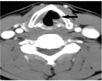
Picture 9. Description CT scan contrast, piece axial, description atrophy flaccid on left vocal fold. Vocal fold in paramedian position 16
Figure 9 is a depiction of a contrast CT scan, axial section, and a atrophy flaccid on plika vocalist left. Plika vocalist on position paramedian. Intrathoracic CT scan can identify intrathoracic causes. MRI examination can also be performed to see abnormalities in the intrathoracic or possible abnormalities in the intracranial. Research conducted by Munin, et al. laryngeal electromyography is a very useful examination to assess vocal fold paralysis and can determine an accurate diagnosis and prognosis in UVFP. Especially if performed 1-6 months after the initial symptoms appear. Examination with this electromyography can identify normal innervation, nerve injury or conduction lesions in the nerves distal, reinnervation And even synkinesis Which rated based on characteristics of nerve electrical signals 16 .
Videostroboscopy is an examination to assess the condition of the larynx by providing a better image and helping with detection. fine movement tape voice, process voice And arytenoid. But examination with videostroboscopy cannot detect mucosal waves. 16 .

Picture 10. Description Videostroboscopy Pre-Operation 17
Figure 10 illustrates the pre-operative videostroboscopy image , in where on picture A And B PPV left during abduction (A) And adduction
(B) there is incomplete closure of the vocal folds during adduction). Picture videostroboscopy post operation (C) moment abduction And (D) moment adduction in patients who have undergone medialization thyroplasty using Montgomery implants silastic. Plika vocalist on position median And increasing closing glottic moment abduction (C) And adduction (D) 17 .
Management Paralysis Plika Vocalist (PPV)
Management Paralysis Plika Vocalist Unilateral (UVFP)
Handling on UVFP aiming For reduce insufficiency glottic and improve voice with speech therapy exercises, surgical procedures, or a combination. both of them. Recovery on UVFP depends on level the damage that happen on nerve Which can in the form of neuropraxia temporary or total nerve damage. Several factors must be considered when determining the best action and treatment tailored to each patient (Table 3).
Table 3. Factor Which Under consideration for Election Management PPV
Patient Factor | Influenceon Treatment (EarlySurgical intervention vs. Observation |
Presence of clinical aspiration | Favors early surgery intervention |
Nature of nerve injury (transection, stretch, orunknown) |
|
Vocal demands of patient |
|
Medical comorbidity |
|
LEMG findings |
|
LEMG: laryngeal electromyography; ML, medialization laryngoplasty; OR, operating room Permanent surgical treatment is performed in UVFP if the cure rate is low due to suspected axonotomesis or neurotomesis, severe persistent dysphonia, aspiration, or lack of response to initial treatment. 14 Treatment options for UVFP are:
Observation And Speech Therapy
Voice therapy is a behavioral treatment designed to reduce the severity of dysphonia and improve functional voice. Characteristics of voice therapy are usually a combination of both direct (behavioral techniques that focus on differences in the voice subsystem) and indirect (advice and guidance to manage external and personal factors that contribute to voice disorders) treatments. Although voice therapy is a common treatment for patients with UVFP, there is limited evidence of its effectiveness, and limited details of the characteristics of effective treatment programs. There is currently no consensus. regarding the content, timing, duration, and frequency of voice therapy delivered as reported for the treatment of other types of voice disorders, such as muscle tension dysphonia . Consequently, there is still great variability in the characteristics of voice therapy which makes it difficult to determine the optimal treatment program for a patient 18 .
Several recent studies on sound therapy for patients with UVFP have show improvement methodology study through implementation treatment protocol And design prospective methodology. Invention time front must focus on development And to design protocol treatment therapy voice, Which as evidence based on Possible, containing exercise special (can replicated) with clearly articulated goals, based on the patient's diagnosis and presenting concerns and needs and containing information about the timing, frequency and duration of treatment 18 .
Randolph points out that there are several different treatment options available. patient Which experiencing PPV temporary, like neuropraxia or gap small glottic (< 1-3 mm). The initial treatment used is speech therapy to overcome dysphonia. This initial therapy aims to prevent muscle atrophy. experience paralysis. Therapy talk aiming For increase vocal fold adduction frequency (target is the non- paralyzed vocal fold) 18 .
In some clinical practices, a combination of speech therapy and surgery is used for the treatment of UVFP. Speech therapy is used before surgery. For minimize hyperfunction focal And therapy talk post surgery to maximize vocal habits. There is limited evidence regarding the effectiveness or success of speech therapy. There is currently no consensus on the timing, duration and frequency of speech therapy that should be performed in patients with UVFP. In some patients who do not recover with conservative therapy, other measures are needed to treat the nerve palsy. 18 .
Injection Laryngoplasty (IL)
Laryngeal injection, also called injection laryngoplasty, has become popular in the last decade. This may be due to several factors, namely: (1). New and safe injection materials; (2). The possibility of using general or local anesthesia; and (3). The choice of several approaches--cricothyroid membrane, thyroid notch , trans-thyroid And cartilage transoral. Proof Which available through meta-analysis showed no significant difference between voice quality outcomes when performed under general or local anesthesia 19 .
Injection laryngoplasty is a technique aimed at augmentation and medialization of the vocal cords to reduce glottic insufficiency caused by various conditions, such as PPV, paresis, or atrophy. IL was first introduced on year 1911 by Bruen as procedure transoral done with anesthesia general. Matter This is technique surgery the oldest Which developed for the management of vocal cord insufficiency. This technique has evolved primarily into procedure based on office. Objective from material Which injected is to match the biomedical and viscoelastic properties of the lamina propria tissue of the vocal folds. Most practitioners choose augmentation injection before considering laryngeal framework surgery 20 .
Indication And Criteria Election Patient on Injection Laryngoplasty (IL)
Injection laryngoplasty may be performed for several conditions that result in glottic insufficiency and altered perception of voice quality. These conditions include PPV, paresis, atrophy, bowing, and scarring. It is important to note that There is Lots condition medical Which can result in development glottic cleft, including thyroid cancer, laryngeal cancer, Parkinson's disease, rheumatoid arthritis, And etc. By Because That, very important for Otolaryngologist to perform a thorough evaluation to diagnose and treat the primary etiology of glottic insufficiency before initiating treatment with IL 20 .
Awake Injection Laryngoplasty (AIL)
Clinicians should be aware that AIL has an initial learning curve that ultimately leads to excellent results with experience. AIL should be the primary technique considered for vocal cord medialization in all patients. However, there are situations where this technique has advantages for maximal outcomes. For example, there is an advantage in performing IL in patients with significant comorbidities that place them at risk for complications related to general anesthesia. Similarly, if the patient have symptoms that No Enough critical in the discomfort and risks of general anesthesia, AIL injection is preferred. Furthermore, awake procedures should be under consideration when There is uncertainty in benefit For injection or when the diagnosis itself is still in question. In these cases, an augmentation trial allow patient For feel level repair sound to make decision information about medialization permanent. Avoid The risks of general anesthesia are particularly relevant in patients requiring secondary augmentation for atrophy, as these patients tend to be older. This is particularly important because IL And surgery laryngoplasty medialization Which can happen produce similar sound results after a 6-month follow-up period 20 .
AIL Also have profit Which allow For evaluation quick from the injection results, based on voice quality, airway patency, location, and diffusion of the injected material. Sulica, et al. showed that continuous visualization of the larynx dynamic allow placement And quantity injection Which optimal to achieve adequate functional results and avoid complications. Furthermore, the use of videostroboscopy before and after injection allows for direct assessment of glottic closure 20 .
There are also cost and time advantages associated with AIL that should be considered. For example, the time required to perform the procedure including consent and administration of topical anesthesia is typically under 30 minutes, eliminating the time required for anesthesia preparation, induction, and operating room preparation. There are also significant cost-effectiveness advantages in reduce cost House Sick And anesthesia. Bove, et al. use predictive model For describe savings Which obtained economy health more than $40 million per year, if there is widespread acceptance and implementation of office-based IL 20 .
- Injection Laryngoplasty Under General Anesthesia
This technique continues to play an important role in allowing for technical control. Which more Good from location needle And distribution injection compared to with injection in an awake patient. Furthermore, the patient is under general anesthesia allowing for additional surgical procedures, such as the removal of autologous fat or fascia. In cases of scarring of the vocal cords, injection in the operating room allow For dissection surgery micro For evaluate more further diagnosis This. Patient worried or No cooperative more Good treated in general anesthesia 20 .
Material On Injection Laryngoplasty
The material used for injection must be biocompatible and inert to reduce the risk of local tissue reactions or fibrosis. The material must also be easy to used And own cost Which low. Besides That, must stand long-lasting and resistant to resorption or migration, while maintaining the normal viscoelasticity of the vocal cords after injection. The materials currently available for injection laryngoplasty are 20 :
Hyaluronic acid-based materials (Restylane, Medicis Aesthetics, Scottsdale, AZ, USA, and Juvederm, Allergan, Irvine, CA, USA)
Autologous fat, carboxymethycellulose (Radiesse Voice Gel, Merz Aesthetics, Inc., Franksville, WI, USA)
Micronized acellular dermal matrix (Cymetra, Life Cell Corp., Branchburg, NJ, USA),
Calcium hydroxylapatite (Radiesse Voice, Merz Aesthetics, Inc., Franksville, WI, USA) [2••].
Older materials, such as Teflon (DuPont, Wilmington, DE, USA), are no longer used because of complications associated with them, such as granuloma formation.
Most of these products were developed as dermal fillers and are not specifically approved for use in the larynx; however, there is widespread acceptance of their use. off-label of the product this is in among otolaryngologists. Because there is no there are materials Which perfect in market, of course so important For understand The advantages and disadvantages of the available materials to select the optimal material for the conditions on the hands. Available materials are as follows 20 :
Carboxymethycellulose (Radiesse Voice Gel, Merz Aesthetics, Inc., Franksville, WI, USA)
Carboxymethylcellulose is made as a gel, consisting of sodium carboxymethylcellulose, glycerin, and water. This material has the advantage of being moist, no allergy testing is required before use, and it is ready to use without the need for preparation or harvesting. This material can last 1-3 months.
Calcium Hydroxylapatite (Radiesse Voice, Merz Aesthetics, Inc., Franksville, Wisconsin, USA)
Collagen Derived Products
This material is a mineral found in human bones and teeth. The injection form is a combination of calcium hydroxylapatite microspheres suspended in a carboxymethylcellu loose carrier gel. Kwon et al. and Rosen, et al. stated that this material can last up to 12 months, while Carrol, et al. showed a mean duration of approximately 18.6 months after injection.
A number of product derivative collagen available For injection. In general historical, material material This engineered from collagen cow or man. Zyplast (Named Aesthetics, Fremont, CA, USA) And Zyderm (McGhan Medical Corp., Fremont, CA, USA) is derived from bovine collagen and lasts up to 4 months. Cosmoplast (Allergan, Irvine, CA, USA) and Cosmoderm (Allergan, Irvine, CA, USA) are derived from human collagen. These materials are no longer available for clinical use. Cymetra (Life Cell Corp., Branchburg, NJ, USA) is not product collagen which is specific, but similar in nature and derived from processed human cadaveric skin tissue. Therefore, it has the potential risk of transmitting pathogens from the tissue. donor, although so far This No There is transmission infection Which reported. Cymetra has a variable average duration from 2 to 3 months to more than 1 year.
Hyaluronic AC ID
This material is a natural polysaccharide that is part of the extracellular matrix. This polysaccharide is also found in the lamina propria of the vocal cords and helps in the repair of their tissue, providing synthetic building blocks for the extracellular matrix. The version of hyaluronic acid available for injection is cross-linked , making it more durable. Rheology studies have shown that hyaluronic acid -based materials have the closest viscoelastic profile resemble tape voice man. Restylane And Hylan b Gel is commercially available preparations used off-label in the larynx are made from cross-linked hyaluronic acid. These materials can last approximately 4–6 months.
Autologous Fat Injection
Autologous Fat Injection has proven give results Which more stand duration of medialization effect after injection compared to other materials. Although the duration has been reported as 26 months, the actual duration varies due to the unpredictable nature of the resorption rate. Fat tissue is usually taken from the subcutaneous tissue of the abdominal wall skin. Because of its autologous nature, this injection is biocompatible and safe to use and a material that is close to the viscoelasticity of the covering layer of vocal cords. One weakness main of injection fat is time development Which long. Besides That, ability endure life fat Which No can predicted cause variability from the results of fat injection. Although there have been reports of donor hematoma, poor voice quality due to over-injection and fat extrusion, the complication rate is low. Another disadvantage of this procedure is the fact that the patient needs to undergo general anesthesia. Patients tend to suffer from prolonged postoperative dysphonia for several weeks due to the need for over-injection.
Table 4. Type Material on Laryngeal Injection 21

UVFP, Unilateral Vocal Paralysis; CaHA, calcium hydroxyapatite; CMC, carboxymethylcellulose; FDA, Food and Drug Administration; PMMA, polymethylmethacrylate
Technique Injection Laryngoplasty (IL)
There are three main approaches to IL, namely: percutaneous, transoral, and microlaryngoscopy.
Percutaneous Injection
IL via percutaneous injection can be performed via the trans-cricothyroid membrane, trans-thyroid cartilage, or trans-thyrohyoid route. The initial steps in preparing for percutaneous injection are:
Localize limitation larynx with palpation. Limitation most important is thyroid notch , limit inferior cartilage thyroid, cartilage cricoid, And membrane cricothyroid.
If wanted, 0.5–1.0 ml local anesthesia can entered with needle inject 1 mL and a 30-gauge needle above the cricothyroid membrane for trans-cricothyroid injection, through the lower border of the thyroid lamina for transthyroid injection, and above the thyroid notch for the trans-thyrohyoid approach.
Injection done in lower visualization direct with help
nasolaryngoscope flexible Which connected to video monitor.
Injection Transcricothyroid
IL through the cricothyroid membrane is the most commonly used approach.
In approach This, membrane cricoid And cricothyroid First time palpated.
The index finger of the non-injecting hand is placed over the cricothyroid membrane. as high as thyroid inferior. Needle injection 27-gauge entered in inferior limit cartilage thyroid, approximately 5 mm lateral to line middle in direction perpendicular to the thyroid.
Then advance it to the tip of the needle contact is made with the cartilage. At this point, the needle tip is advanced inferiorly to guide the tip beneath the inferior border of the thyroid cartilage.
Important For use tactile bait come back from needle in lower limit thyroid, as it will confirm that the needle will go into the paraglottic space. With end needle in position inside, make sure For No interesting back to the hand syringe, then it can be used to make a bend in the needle to a 30 angle at the hub.
It is done by pushing the syringe into the skin of the neck while the tip of the needle points superiorly and lateral. At this point, the needle slowly proceed 5– 10 mm in position This, going to superior And lateral. While see in video monitor, the needle is gently manipulated while injecting a small amount of additional material to help identify the position of the intralaryngeal tip.
The material is injected while visualizing for adequate medialization of the vocal cords.
Transthyroid Cartilage Approach
For this approach, the borders of the larynx are also palpated, and the index finger is placed at the level of the cricothyroid membrane.
Needle placed upright straight to cartilage thyroid, approximately 5 mm lateral to the line middle And 2-3 mm in on limit inferior. As level plika vocalist usually located in middle between thyroid notch And limit inferior thyroid cartilage, If limitation can touched, somebody can make description mental about the estimated injection target.
Needle Then Can placed on level vertical in point middle between the superior vocal cords and the inferior border of the thyroid cartilage.
Gently press until the needle enters the cartilage. At this point, the needle is bent slightly at the hub to point laterally toward the paraglottic space.
Injection Then can done while observe mediation on video monitor
Transhyoid Approach
After local anesthesia infiltration above the thyroid notch and thyrohyoid membrane area, a syringe filled with 4% lidocaine is injected through the thyrohyoid membrane into the airway.
Next, under direct visualization with a nasolaryngoscope , a syringe filled with injection material is attached to the appropriate needle.
The needle is inserted just above the thyrohyoid notch and through the subcutaneous tissue below the angle, passing through the pre-epiglottic space and entering the larynx at the level of the epiglottis stalk. Next, the needle is slowly advanced to the desired injection site unilaterally or bilaterally.
Transoral Approach
This approach can be performed in an operating room or office setting under direct visualization with the aid of a nasolaryngoscope .
First, tongue the patient is held in place with gauze one hand while the syringe with the injection material is held in the other hand. Usually a 25 cm long needle with a 16 gauge soft shaft and a 25 cm needle tip is used for injection.
The needle is bent precisely so that it can be guided through the pharynx and into the larynx. While the assistant holds the flexible nasolaryngoscope, the needle is inserted orally and guided toward the appropriate vocal fold. The ingredients must injected lateral to tape voice on level vocal process .
Transnasal, Endoscopy
This approach can be performed in an office setting under topical anesthesia. The flexible telescope with working method is introduced transnasally at the level of the laryngopharynx.
Anesthesia larynx obtained with application topical solution lidocaine 4%.
The sclerotherapy needle is filled with injection material and then passed through the channel. Work telescope And directed to mucosal to in room paraglottic lateral to the affected vocal cord for augmentation injection. This technique is limited by the relatively long length of the injection tube which increases resistance to flow material moment inject, so that it requires a large amount enough pressure injection For push out material.
Materials of higher viscosity cannot be injected with this technique, but it has been used successfully with many materials used in the office setting.
Microlarygoscope Approach
Approach This done with anesthesia general And in method Which same as the transoral approach.
After the patient is intubated, head place Sleep played 90 from the anesthesiologist .
Larynx open And suspended with use laryngoscopy Which rigid.
Using an operating microscope to visualize the vocal cords, a 25-cm long, soft-tipped 25-gauge needle is guided through a rigid laryngoscope. As in the transoral approach, the needle is guided laterally to the vocal cords at the level of the vocal processes.
Operation Laryngeal Skeleton
Surgical intervention is recommended in patients in whom injection augmentation therapy has proven to be of no benefit, in cases of obvious aspiration due to vocal fold paralysis or in whom there is evidence of severe denervation injury on examination. laryngeal electromyography. Surgical intervention Which can be done is medialization thyroplasty.
Technique thyroplasty medialization introduced by isshiki, action This performed by making a hole in the thyroid cartilage by inserting a permanent alloplastic implant for medialization of the vocal folds. This technique is considered the gold standard for the treatment of vocal fold paralysis. This technique is reported to provide excellent results by providing improvements in voice quality. Commonly used alloplastic materials include silicone silastic polymer, polytetrafluoroethylene (gore-Tex), titanium, and montgomery implants. The advantages of this technique are lower cost and a relatively permanent technique even though the implant can be removed surgically. Rare complications of this procedure can be edema and hyperemia 16 .
Besides thyroplasty medialization, action surgery Which can done for the treatment of PPV is arytenoid adduction. This technique was first introduced by Ishiiki in 1978. The purpose of arytenoid cartilage adduction is to reduce large glottic incompetence, especially in the posterior part. This technique this is done with how to use thread Which missed around the muscular process of the arytenoids through the anterior thyroid cartilages, then pulled with sufficient tension to adduct the arytenoids to the midline so that they cover commissure posterior. Action This can increase complications in the form of dysphagia and airway obstruction due to laryngeal edema compared with surgical intervention medialization thyroplasty. Technique This arytenoid adduction is often combined with medialization thyroplasty or injection laryngoplasty 16 .
Management Paralysis Plika Vocalist Bilateral (VFPB)
Surgery to treat VFPB has evolved over the past few decades. Moment This endoscopy transoral or microscopic surgery give results the better in many cases compared to external surgical procedures that damage the laryngeal framework. Arytenoidectomy, posterior cordectomy, and laterofixation are standard treatment procedures for VFPB. VFPB can cause life-threatening airway obstruction and often requires emergency surgery. In these cases, airway patency is the primary concern. Various procedures that can be carried out in the management of VFPB are as follows. 22
Tracheostomy
It is one of the most common procedures used in cases of VFPB. Tracheostomy provides a direct bypass from the airway obstruction. Despite being a very effective method, tracheostomy is not preferred by most patients, due to the presence of an open wound, the need for long-term care, and decreased quality of life. Naunheim et al reported that endoscopic techniques are cheaper than tracheostomy in the treatment of VFPB. However, in cases of sudden onset and severe dyspnoea , tracheostomy remains the gold standard in patients with PPV.
Arytenoidectomy
Depending on its function, the rima glottis may be divided into a phonatory segment (anterior 2/3) and a respiratory segment (posterior 1/3). The arytenoid cartilages are the main keys of the posterior part of the larynx. Arytenoidectomy (removal of the arytenoid cartilages) is an irreversible surgical method to widen the rima glottis in axis transversal. Technique This applied expand in a way independent or in combination with resection partial plika vocalist. Arytenoidectomy First done in 1916 by Baker, which in a way simultaneously Partial cordectomy was performed through the laryngofissure approach. Iwanoff removed the arytenoid cartilage by incising the mucous membrane in the medial laryngeal line, while Kelly created a window in the inferoposterior aspect of the thyroid cartilage. Other approaches are described by Woodman, Which incise skin in a way parallel to edge front sternocleidomastoid muscle at the level of the superior border of the thyroid cartilage through the cricothyroid joint posterior. Approach This make it easier maintenance plate the thyroid cartilage as a whole, and the remaining vocal process is sutured laterally inferiorly to the thyroid cartilage.
More effective lateralization and smaller vertical displacement were obtained by Newman et al. who modified the above technique. The vocal process was attached through a small hole in the thyroid cartilage at the level of the cricoarytenoid joint. Helmus described arytenoidectomy through a thyreofissure (only the thyroid cartilage is divided) under microscopic conditions with the use of microinstruments that allow dissection Which appropriate from cartilage arytenoids. Arytenoidectomy which is accessed via a medial thyrotomy performed by Perlman and Kilian, who removed the cartilage through a small incision in the mucous membrane medially. They use electrocoagulation, Which make it easier achieving hemostasis and causing the development of scar tissue that results in a lateral position from the vocal cords.
On year 1948, Thornell describe arytenoidectomy intra-laryngeal with an endoscopic approach. After a temporary tracheotomy and injecting the vocal folds and folds aryepiglottic with anesthetic solution, an incision is made in above the arytenoid cartilages as far as the aryepiglottic folds. The cartilages are dissected and removed through the access obtained. Additional tissue lateralization is obtained with the use of electrocoagulation And formation network scar. A stent Acrylic is placed between the vocal cords to cut the vocal folds, vocal ligaments, or thyroarytenoid muscles.
Cordectomy
Cordectomy (posterior And transversal) is method irreversible from widening of the rima glottis involving the removal of part of the vocal folds, vocal ligaments, or thyroarytenoid muscles. Cordectomy was first described by Hoope in 1896, who performed a simultaneous tracheotomy on the patient. The operation did not produce the anticipated effects. Several years later, in 1908, Citelli described an external cordectomy performed through a thyreofissure approach. Hoover presented Jackson's technique called "ventriculochordectomy", in which the vocal folds (forming the inferior part of the ventricle of the larynx - referred to as "ventriculo" by the author) were removed through a laryngofissure approach, the vocal ligaments ("cordectomy"), and the vocal processes. Surjan modified the cordectomy performed through a thyreofissure. He created a flap of mucous membrane in the larynx - at the level of the vestibular folds and contralateral other - in subglottis. Innovation like that aiming reduce risk of stenosis. Šercer reported total bilateral submucosal resection of the vestibular and vocal folds together with the anterior part of the arytenoid cartilages. The author claims that only part posterior from cartilage arytenoid maintained on cartilage cricoid in "neolarynx". He observe voice hoarse on patient, produced by the aryepiglottic folds.
After endoscopic laser surgery was performed in the treatment of VFPB, Dennis and Kashima presented their technique in 1989. They used a CO2 laser to cut a C-shaped section of cartilage from the plica vocalist in part posterior with transsection elastic cone . They achieved decannulation in 60% of patients, but bilateral or repeat cordectomy was required in 30% of them. The technique presented by Dennis and Kashima has been modified by Pia And Banana. Plika vocalist Which restored widen to sideways and backwards along the vestibular fold with a CO2 laser. The mucous membrane lesion is closed with fibrin preparation. Cordectomy front submucosa is done by Ekel. Cartilage arytenoid intact, And folds membrane mucus closed with fibrin glue to intralarynx lesions. The results obtained are consistent with laser arytenoidectomy. conventional. Benninger, et al. Also display laser cordectomy posterior with resection submucosa process vocalist And formation mucous membrane, which is then sutured laterally to the created pocket.
Laterofixation
It is Wrong One action Which can done For open road breath in patients with bilateral vocal fold paralysis. This technique was popularized by Lichtenberger and Toohill. This technique uses non-absorbable sutures placed endoscopically above and below the posterior vocal cords. This can applied in a way independent or in combination with other laryngeal methods unilateral or bilateral microsurgery. The rima glottis is widened without damage network For phonation. According to reviews by Sapundzhiev, et al. fixation of the cricoarytenoid joint by laryngofissure was first described. by Rethy on year 1922. He use stent intra larynx For 2- 3 month post operation For maintain mobilization cartilage arytenoid laterally. A similar technique was used by Montgomery who applied a special stainless-steel pin technique .
Lateralization endoscopic with stitches done by Kirchner on year 1979 and Ejnell in 1982. Kirchner performed endoscopic resection of the thyroarytenoid muscle with electrocoagulation. He temporarily repaired the vocal folds with sutures inserted into the laryngeal lumen. Ejnell et al. performed lateralization of the vocal folds as an independent procedure. They visualized the larynx through direct laryngoscopy and identifying the cricoarytenoid joints. With use two needle Which entered in a way externally, they tied the knot and lateralized the vocal folds. Remsel modified Kirchner's technique by replacing electrocoagulation with CO2 laser.
Endo-extralaryngeal access in laterofixation with the use of a specially developed needle was introduced by Lichtenberger, in which the sutures are placed from lumen larynx, through the lamina cartilage thyroid, and out to the skin. He also classified laterofixation into reversible endo-extralaryngeal, endo-extralaryngeal lateral laryngo-microsurgical lateralization and laryngo- microsurgical endo-extralaryngeal with arytenoidectomy. In the beginning resection performed with surgical instruments, which were later replaced by CO2 laser. A modification of the above technique was proposed by Woodson and Weiss. Lateralization with sutures was performed at the level of the arytenoid cartilages . They called it " arytenoid abduction ", and caused postero-caudal traction that stimulated contraction of the posterior cricoarytenoid muscle.
Prognosis Paralysis Plika Vocalist (PPV)
The results of PPV therapy are very good. Most patients can return to almost normal or even normal speech and with minimal or no limitation of speech function for daily speaking needs, but for singing, it is likely not possible to be perfect, because the ability of the vocal cords is already limited 12 .
Complications Paralysis Plika Vocalist (PPV)
Complications from therapy surgery is voice Which not enough Good, difficulty breathing, and migration of the implant. At the time of surgery which includes manipulation from channel breath, factor like hematoma, edema can cause trouble breathe, And For prevent from complications This so on moment The operation must be carried out precisely and very carefully, as well as with the administration of pre- and post-operative corticosteroids, and the risk will be greater if the surgical process is bilateral.
Although surgery is essential if there is dysphagia, most surgeries are performed to improve voice quality, and if there is no improvement in voice quality, then complications occur during the procedure. Often poor voice quality or No there is a fix after surgery can corrected by repeat medialization laryngoplasty with or without arytenoid adduction and the most common cause of poor voice quality after surgery is misplacement of the implant, placement too anterior/superior, implant too small/large. This can cause intraoperative edema, which can be prevented by the use of corticosteroids to minimize edema before can done return replacement implant. Migration of the implant may occur post-operatively, either medially into the airway or laterally into the neck.
DISCUSSION
Complaints of voice disorders are not uncommon to find in clinics. Voice disorders or dysphonia can be in the form of a hoarse voice, which is a voice that sounds rough with a lower tone than usual, a weak voice, loss of voice, a tense voice that is difficult to get out, a voice consisting of several tones, pain when making a sound or the inability to reach a certain tone or intensity. Phonation or sound production is a function of the larynx that is produced by the tension of the vocal cords. The work of the vocal cords is produced from the movement intrinsic muscles Which is in the larynx. If there is a disturbance in muscle function due to an abnormality in the muscles the or dysfunction nerve can cause the disturbance production sounds like the complaint above.
A retrospective study at Taipei Hospital of medical records of patients with PPV from September 2010 to December 2019 was conducted to determine the etiology of vocal fold paralysis. Patients with laryngeal/hypopharyngeal malignancy, in whom examination and follow-up data were incomplete, were excluded from the study. During act carry on, case Which involving recovery Also issued. Vocal cord paralysis can be caused by various diseases or surgery. There are many studies on PPV, but the incidence of PPV is still difficult to determine. This study showed that the incidence of PPV increases with the age of the patient. The peak incidence is between 51 and 70 years which may be due to the increased incidence of tumors and neurological damage in aging individuals. More male patients than females have unilateral or bilateral PPV, probably because there is an increase in postoperative cases acquired from lung surgery and esophageal cancer in men 3 .
Research by Lucian Lusica et al was conducted on 34 patients treated with injection medialization. laryngoplasty through a percutaneous approach. Other approaches used for the procedure include: peroral, percutaneous transthyrohyoid and percutaneous transthyroid cartilage route . They use agarose and hyaluronic acid as tissue fillers for injection into patients. Both of these materials are tissue fillers that have been established for human use. The ingredients This weak And can injected in a way percutaneous. Loss from material- This material is that it tends to be absorbed with time. All patients had significant voice improvement as assessed by voice analysis using different subjective and objective parameters. Collagen, fat, and Teflon are other materials commonly used for this procedure. From Unlike the above agents, only fat and Teflon can be injected percutaneously, whereas collagen can only be injected orally under direct laryngoscopic control 3 .
The static glottic enlargement procedure consists of removing laryngeal tissue (arytenoidectomy, cordectomy) or shift structure anatomy (laterofixation vocal folds). In addition, laterofixation may be considered as an alternative to tracheostomy, due to its reversibility and minimal damage to vocal and swallowing function. Other techniques such as Botox injection, reinnervation, or laryngeal stimulation, are indicated. on patient with VFPB, in where synkinesis larynx observed. The technique involves restoring physiological function of the larynx without performing tissue resection. Therapy gene And therapy stem cells currently in stage test on animal. They aim to encourage the regrowth of neurons and atrophic muscle cells. modality depends on duration VFPB. Reinnervation larynx including end-to-end anastomosis in cases of nerve arrest is performed during the first six months after bilateral iatrogenic injury to the RLN. Laser posterior cordectomy combined with partial arytenoidectomy is selected in cases performed if more than six months have passed since then diagnosis of VFPB 22 .
CONCLUSION
Vocal cord paralysis is a loss of movement or paralysis of the vocal cords. This condition has a profound impact on the quality of life of sufferers and is an important issue in the field of otolaryngology. Condition This is manifestation from disease Which happen in the cranial cavity, larynx, thorax and mediastinum causing paralysis of the vagus nerve and recurrent laryngeal nerve. Unilateral vocal fold paralysis (UVFP) or bilateral (VFPB) can interfere with the physiological function of the larynx, namely breathing, protection of the airway and phonation. Vocal fold paralysis can occur due to mechanical fixation or due to neurogenic disorders. 4
The symptoms that arise from vocal fold paralysis depend on the damage to the larynx and the symptoms that arise vary from mild to severe. heavy And can nature damage unilateral or bilateral. Paralysis vocal cords can occur in children or adults. This is because the damage to the nerves is permanent. Various interventions have been developed to minimize the damage that occurs.
Some cases of unilateral vocal fold paralysis do not require surgical intervention. Temporary augmentation injections are very effective in reducing symptoms and improving quality of life. However, it does not reduce the possibility of requiring surgical intervention in patients with unilateral vocal fold paralysis. The mainstay of treatment for bilateral vocal fold paralysis is to clear the airway and Sometimes a tracheostomy is necessary immediately to overcome airway obstruction.
REFERENCE
Al-Khtoum, N., Shawakfeh, N., Al-Safadi, E., Al-Momani, O. & Hamasha, K. Acquired unilateral vocal fold paralysis: Retrospective analysis of a single institutional experience. N Am J Med Sci 5 , 699–702 (2013).
Lalwani, AK CURRENT Diagnosis and Treatment in Otolaryngology-- Head and Neck Surgery: Second Edition (LANGE CURRENT Series) . (2008).
Wang, HW, Lu, CC, Chao, PZ & Lee, FP Causes of Vocal Fold Paralysis. Ear Nose Throat J 101 , NP294–NP298 (2022).
Adams, G. L., Boies, L. R. & Gighler, P. A. BOIES Book Teach Disease ENT . ECG (1997).
Hansen, JT & Lambert, DR In Netter's Clinical Anatomy . (Medimedia, 2005).
Witterick, I. Annual Report Annual Report 2018/2019. Fresenius.Com
Snow, J. B. & Ballenger, J. J. In Ballenger's Otorhinolaryngology Head And Neck Surgery . (BC Decker Inc, 2003).
Dance, J. & Jr., M. Anatomy and Physiology of The Voice. (2011).
Dhillon, RS & East, CA In Ear, Nose, and Throat and Head and Neck Surgery . (Harcourt, 2000).
Thomas R. Van With Water, Hinrich, S. In Otolaryngology Basic Science And Clinical Review . (Thieme Medical Publishers, 2005).
Wang, H. W., You, C. C., Chao, P. Z. & Lee, F. P. Causes of Vocal Fold Paralysis. Ear Nose Throat J 101 , (2022).
Al-Khtoum, N., Shawakfeh, N., Al-Safadi, E., Al-Momani, O. & Hamasha Again, I. I. A. & Marie, J. ARTICLE IN PRESS Early Management of Acute Unilateral Vocal Fold Paralysis : Update of the Literature. 19–21 (2020) doi:10.1016/j.jvoice.2020.02.021.
Efianty a., Nurbaity Iskandar, JBRD (2007). D. Textbook of ENT Health Sciences . (FKUI, 2007).
Janet W, et al. Bilateral Selective Laryngeal Reinnervation for Bilateral Vocal Fold Paralysis in Children. 1 , 1–7 (2020).
Siu, J., Tam, S. & Fung, K. A comparison of outcomes in interventions for unilateral vocal fold paralysis: A systematic review. Laryngoscope 126 , 1616–1624 (2016).
Kumar, V. & Lewin LE., G. CT Assessment Of Vocal Cord Medializatiion. (2006).
Walton, C., Carding, P. & Flanagan, K. Perspectives on voice treatment for unilateral vocal fold paralysis. Curr Opin Otolaryngol Head Neck Surg 26 , 157–161 (2018).
Sant'Anna, GD Immediate and early injection in unilateral vocal fold paralysis. Braz J Otorhinolaryngol 86 , 1–2 (2020).
Salinas, J. B. & Chhetri, D. K. Injection Laryngoplasty: Techniques and (2019). K. Acquired unilateral vocal fold paralysis: Retrospective analysis of a single institutional experience. N Am J Med Sci 5 , 699–702 (2013). Choices of Fillers. Curr Otorhinolaryngol Rep 2 , 131–136 (2014).
Ryu, CH , et al. Guidelines for the management of unilateral vocal fold paralysis from the Korean society of laryngology, phoniatrics and logopedics. Clin Exp Otorhinolaryngol 13 , 340–360 (2020).
Czesak, M. A., Osuch-wojcikiewicz, E. & Niemczyk, K. Methods of surgical treatment of bilateral vocal fold paralysis. 350–358 (2020) doi:10.5603/EP.
Comments